
| 产品编号 | bsm-52053R |
| 英文名称 | Cytokeratin 13 Recombinant Rabbit mAb |
| 中文名称 | 细胞角蛋白13重组兔单抗 |
| 别 名 | CK13; K13; WSN2; Krt-1.13; Krt1-13; K1C13_HUMAN; KRT13; Cytokeratin-13 (CK-13); Keratin-13 (K13); K1C13_MOUSE; 47 kDa cytokeratin; |
| 研究领域 | 信号转导 细胞类型标志物 |
| 抗体来源 | Rabbit |
| 克隆类型 | Recombinant |
| 克 隆 号 | 5A3 |
| 交叉反应 | Human,Mouse |
| 产品应用 | WB=1:500-2000,IHC-P=1:100-500,IHC-F=1:100-500,IF=1:100-500,ICC/IF=1:50-200
not yet tested in other applications. optimal dilutions/concentrations should be determined by the end user. |
| 理论分子量 | 49 kDa |
| 检测分子量 | 49 |
| 细胞定位 | 细胞浆 |
| 性 状 | Liquid |
| 浓 度 | 1mg/ml |
| 免 疫 原 | A synthesized peptide derived from human Cytokeratin 13: 121-155/458 |
| 亚 型 | IgG |
| 纯化方法 | affinity purified by Protein A |
| 缓 冲 液 | 0.01M TBS (pH7.4) with 1% BSA, 0.02% Proclin300 and 50% Glycerol. |
| 保存条件 | Shipped at 4℃. Store at -20℃ for one year. Avoid repeated freeze/thaw cycles. |
| 注意事项 | This product as supplied is intended for research use only, not for use in human, therapeutic or diagnostic applications. |
| PubMed | PubMed |
| 产品介绍 |
The protein encoded by this gene is a member of the keratin gene family. The keratins are intermediate filament proteins responsible for the structural integrity of epithelial cells and are subdivided into cytokeratins and hair keratins. Most of the type I cytokeratins consist of acidic proteins which are arranged in pairs of heterotypic keratin chains. This type I cytokeratin is paired with keratin 4 and expressed in the suprabasal layers of non-cornified stratified epithelia. Mutations in this gene and keratin 4 have been associated with the autosomal dominant disorder White Sponge Nevus. The type I cytokeratins are clustered in a region of chromosome 17q21.2. Alternative splicing of this gene results in multiple transcript variants; however, not all variants have been described. [provided by RefSeq, Jul 2008]. Subunit: Heterotetramer of two type I and two type II keratins. keratin-13 is generally associated with keratin-4. Tissue Specificity: Defects in KRT13 are a cause of white sponge nevus of cannon (WSN) . WSN is a rare autosomal dominant disorder which predominantly affects non-cornified stratified squamous epithelia. Clinically, it is characterized by the presence of soft, white, and spongy plaques in the oral mucosa. The characteristic histopathologic features are epithelial thickening, parakeratosis, and vacuolization of the suprabasal layer of oral epithelial keratinocytes. Less frequently the mucous membranes of the nose, esophagus, genitalia and rectum are involved. DISEASE: White sponge nevus of cannon (WSN) [MIM:193900]: Rare autosomal dominant disorder which predominantly affects non-cornified stratified squamous epithelia. Clinically, it is characterized by the presence of soft, white, and spongy plaques in the oral mucosa. The characteristic histopathologic features are epithelial thickening, parakeratosis, and vacuolization of the suprabasal layer of oral epithelial keratinocytes. Less frequently the mucous membranes of the nose, esophagus, genitalia and rectum are involved. Note=The disease is caused by mutations affecting the gene represented in this entry. Similarity: Belongs to the intermediate filament family. SWISS: P13646 Gene ID: 3860 Database links: Entrez Gene: 3860 Human Entrez Gene: 16663 Mouse Omim: 148065 Human SwissProt: P13646 Human SwissProt: P08730 Mouse Unigene: 654550 Human Unigene: 4646 Mouse |
| 产品图片 |

25 ug total protein per lane of various lysates (see on figure) probed with Cytokeratin 13 monoclonal antibody, unconjugated (bsm-52053R) at 1:1000 dilution and 4°C overnight incubation. Followed by conjugated secondary antibody incubation at r.t. for 60 min.
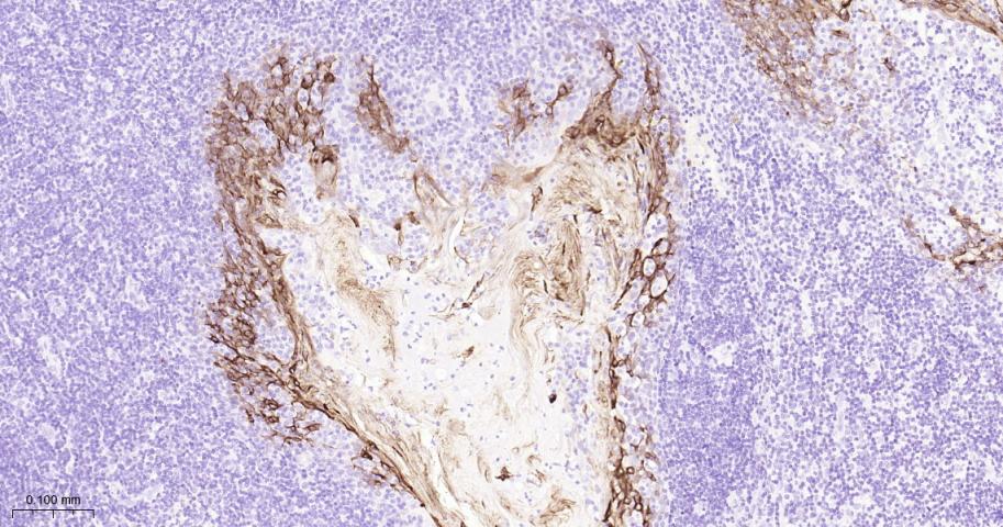
Paraformaldehyde-fixed, paraffin embedded Human Tonsil; Antigen retrieval by boiling in sodium citrate buffer (pH6.0) for 15 min; Antibody incubation with Cytokeratin 13 Monoclonal Antibody, Unconjugated(bsm-52053R) at 1:200 overnight at 4°C, followed by conjugation to the bs-0295G-HRP and DAB (C-0010) staining.

Paraformaldehyde-fixed, paraffin embedded Human bladder invasive urothelial carcinoma; Antigen retrieval by boiling in sodium citrate buffer (pH6.0) for 15 min; Antibody incubation with Cytokeratin 13 Monoclonal Antibody, Unconjugated(bsm-52053R) at 1:200 overnight at 4°C, followed by conjugation to the bs-0295G-HRP and DAB (C-0010) staining.

Paraformaldehyde-fixed, paraffin embedded Human Bladder; Antigen retrieval by boiling in sodium citrate buffer (pH6.0) for 15 min; Antibody incubation with Cytokeratin 13 Monoclonal Antibody, Unconjugated(bsm-52053R) at 1:200 overnight at 4°C, followed by conjugation to the bs-0295G-HRP and DAB (C-0010) staining.

(Negative control) Paraformaldehyde-fixed, paraffin embedded Human Glioma; Antigen retrieval by boiling in sodium citrate buffer (pH6.0) for 15 min; Antibody incubation with Cytokeratin 13 Monoclonal Antibody, Unconjugated(bsm-52053R) at 1:200 overnight at 4°C, followed by conjugation to the bs-0295G-HRP and DAB (C-0010) staining.

Paraformaldehyde-fixed, paraffin embedded Human Esophagus; Antigen retrieval by boiling in sodium citrate buffer (pH6.0) for 15 min; Antibody incubation with Cytokeratin 13 Monoclonal Antibody, Unconjugated(bsm-52053R) at 1:200 overnight at 4°C, followed by conjugation to the bs-0295G-HRP and DAB (C-0010) staining.

Paraformaldehyde-fixed, paraffin embedded Mouse Esophagus; Antigen retrieval by boiling in sodium citrate buffer (pH6.0) for 15 min; Antibody incubation with Cytokeratin 13 Monoclonal Antibody, Unconjugated(bsm-52053R) at 1:200 overnight at 4°C, followed by conjugation to the bs-0295G-HRP and DAB (C-0010) staining.
|
| 1、抗体溶解方法 | |
| 2、抗体修复方式 | |
| 3、常用试剂的配制 | |
| 4、免疫组化操作步骤 | |
| 5、免疫组化问题解答 | |
| 6、Western Blotting 操作步骤 | |
| 7、Western Blotting 问题解答 | |
| 8、关于肽链的设计 | |
| 9、多肽的溶解与保存 | |
| 10、酶标抗体效价测定程序 | |


