
| 产品编号 | bsm-52088R |
| 英文名称 | HDAC8 Recombinant Rabbit mAb |
| 中文名称 | 组蛋白去乙酰化酶8重组兔单抗 |
| 别 名 | CDA07; CDLS5; HD8; HDACL1; KDAC8; MRXS6; RPD3; WTS; 2610007D20Rik; RGD1562895; HDAC8_HUMAN; HDAC8; Protein deacetylase HDAC8; Protein decrotonylase HDAC8; 3.5.1.98; HDAC8_MOUSE; HDAC8_RAT; |
| 研究领域 | 肿瘤 发育生物学 信号转导 细胞凋亡 转录调节因子 表观遗传学 |
| 抗体来源 | Rabbit |
| 克隆类型 | Recombinant |
| 克 隆 号 | 4C3 |
| 交叉反应 | Human |
| 产品应用 | WB=1:500-2000,Flow-Cyt=1:50-100,ICC/IF=1:50-200
not yet tested in other applications. optimal dilutions/concentrations should be determined by the end user. |
| 理论分子量 | 42 kDa |
| 检测分子量 | 42 |
| 细胞定位 | 细胞核 细胞浆 |
| 性 状 | Liquid |
| 浓 度 | 1mg/ml |
| 免 疫 原 | A synthesized peptide derived from human HDAC8: 50-110 |
| 亚 型 | IgG |
| 纯化方法 | affinity purified by Protein A |
| 缓 冲 液 | 0.01M TBS (pH7.4) with 1% BSA, 0.02% Proclin300 and 50% Glycerol. |
| 保存条件 | Shipped at 4℃. Store at -20℃ for one year. Avoid repeated freeze/thaw cycles. |
| 注意事项 | This product as supplied is intended for research use only, not for use in human, therapeutic or diagnostic applications. |
| PubMed | PubMed |
| 产品介绍 |
Histones play a critical role in transcriptional regulation, cell cycle progression, and developmental events. Histone acetylation/deacetylation alters chromosome structure and affects transcription factor access to DNA. The protein encoded by this gene belongs to class I of the histone deacetylase family. It catalyzes the deacetylation of lysine residues in the histone N-terminal tails and represses transcription in large multiprotein complexes with transcriptional co-repressors. Multiple transcript variants encoding different isoforms have been found for this gene. [provided by RefSeq, Oct 2009]. Function: Responsible for the deacetylation of lysine residues on the N-terminal part of the core histones (H2A, H2B, H3 and H4). Histone deacetylation gives a tag for epigenetic repression and plays an important role in transcriptional regulation, cell cycle progression and developmental events. Histone deacetylases act via the formation of large multiprotein complexes. May play a role in smooth muscle cell contractility. Subunit: Interacts with PEPB2-MYH11, a fusion protein consisting of the 165 N-terminal residues of CBF-beta (PEPB2) with the tail region of MYH11 produced by the inversion Inv(16)(p13q22), a translocation associated with acute myeloid leukemia of M4EO subtype. The PEPB2-MYH1 fusion protein also interacts with RUNX1, a well known transcriptional regulator, suggesting that the interaction with HDAC8 may participate in the conversion of RUNX1 into a constitutive transcriptional repressor. Interacts with CBFA2T3. Interacts with phosphorylated SMG5/EST1B; this interaction protects SMG5 from ubiquitin-mediated degradation. Associates with alpha-SMA (smooth muscle alpha-actin). Subcellular Location: Nucleus. Cytoplasm. Excluded from the nucleoli. Found in the cytoplasm of cells showing smooth muscle differentiation. Tissue Specificity: Weakly expressed in most tissues. Expressed at higher level in heart, brain, kidney and pancreas and also in liver, lung, placenta, prostate and kidney. Post-translational modifications: Phosphorylated by PKA on serine 39. Phosphorylation reduces deacetylase activity observed preferentially on histones H3 and H4. Similarity: Belongs to the histone deacetylase family. HD type 1 subfamily. SWISS: Q9BY41 Gene ID: 55869 Database links: Entrez Gene: 55869 Human Entrez Gene: 70315 Mouse Omim: 300269 Human SwissProt: Q9BY41 Human SwissProt: Q8VH37 Mouse Unigene: 310536 Human Unigene: 328128 Mouse Unigene: 208476 Rat |
| 产品图片 |
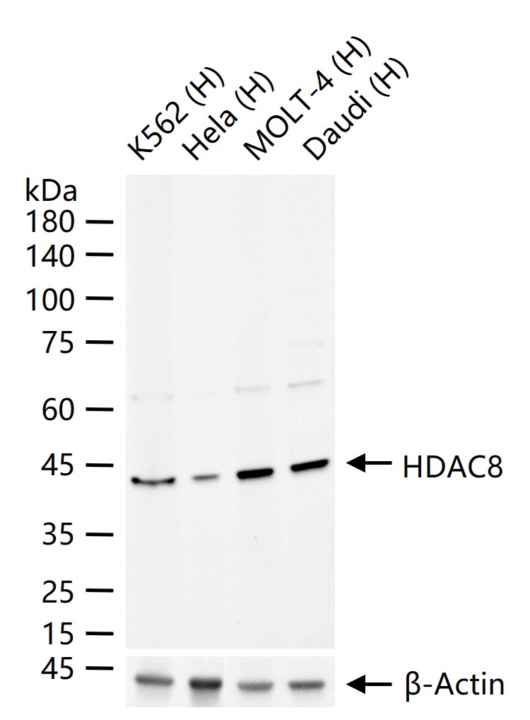
25 ug total protein per lane of various lysates (see on figure) probed with HDAC8 monoclonal antibody, unconjugated (bsm-52088R) at 1:1000 dilution and 4°C overnight incubation. Followed by conjugated secondary antibody incubation at r.t. for 60 min.
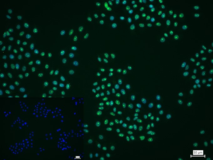
4% Paraformaldehyde-fixed HepG2 (H) cell; Triton X-100 at r.t. for 20 min; Antibody incubation with (HDAC8) monoclonal Antibody, unconjugated (bsm-52088R) 1:200, 90 min at 37°C; followed by conjugated Goat Anti-Rabbit IgG antibody (green, bs-60295G-BF488) at 37°C for 90 min, DAPI (blue, C02-04002) was used to stain the cell nuclei. PBS instead of the primary antibody was used as the blank control.
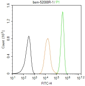
The HepG2 (H) cells were fixed with 4% PFA (10 min at r.t.) and then permeabilized with 90% ice-cold methanol for 20 min at -20℃,the cells then were incubated in 5%BSA to block non-specific protein-protein interactions (30 min at r.t.).Primary Antibody (green):Rabbit Anti-HDAC8 antibody (bsm-52088R): 1 μg/10^6 cells; Secondary Antibody : Goat anti-Rabbit IgG-BF488 (bs-60295G-BF488): 1 μg/test. Isotype Control (orange): Rabbit IgG (bs-0295P). Blank control (black): PBS. Acquisition of 20,000 events was performed.
|
| 1、抗体溶解方法 | |
| 2、抗体修复方式 | |
| 3、常用试剂的配制 | |
| 4、免疫组化操作步骤 | |
| 5、免疫组化问题解答 | |
| 6、Western Blotting 操作步骤 | |
| 7、Western Blotting 问题解答 | |
| 8、关于肽链的设计 | |
| 9、多肽的溶解与保存 | |
| 10、酶标抗体效价测定程序 | |


