
| 产品编号 | bsm-54170R |
| 英文名称 | PSMA1 Recombinant Rabbit mAb |
| 中文名称 | 蛋白酶体PSMα1重组兔单抗 |
| 别 名 | HC2; HEL-S-275; NU; PROS30; C2; Pros-30; alpha-type; PSA1_CHICK; PSMA1; Macropain subunit C2; Multicatalytic endopeptidase complex subunit C2; Proteasome component C2; PSA1_HUMAN; 30 kDa prosomal protein (PROS-30); Proteasome nu chain; Proteasome subunit |
| 研究领域 | 细胞生物 免疫学 |
| 抗体来源 | Rabbit |
| 克隆类型 | Recombinant |
| 克 隆 号 | 10A2 |
| 交叉反应 | Human,Mouse,Rat |
| 产品应用 | WB=1:500-2000,IHC-P=1:50-200,IHC-F=1:50-200,IF=1:50-200,Flow-Cyt=1:50-200,ICC/IF=1:50-200
not yet tested in other applications. optimal dilutions/concentrations should be determined by the end user. |
| 理论分子量 | 30 kDa |
| 检测分子量 | 32 |
| 细胞定位 | 细胞核 细胞浆 |
| 性 状 | Liquid |
| 浓 度 | 1mg/ml |
| 免 疫 原 | Recombinant human PSMA1 |
| 亚 型 | IgG |
| 纯化方法 | affinity purified by Protein A |
| 缓 冲 液 | 0.01M TBS (pH7.4) with 1% BSA, 0.02% Proclin300 and 50% Glycerol. |
| 保存条件 | Shipped at 4℃. Store at -20℃ for one year. Avoid repeated freeze/thaw cycles. |
| 注意事项 | This product as supplied is intended for research use only, not for use in human, therapeutic or diagnostic applications. |
| PubMed | PubMed |
| 产品介绍 |
Ubiquitin-dependent proteolysis mediates selective destruction of various cell cycle regulators, transcription factors and tumor suppressors. In eukaryotic cells, selective breakdown of cellular proteins is ensured by their ubiquitination and subsequent degradation by the 26S proteasome. At specific stages of development, embryo- and tissue-specific components of the 26S proteasome form, facilitating proteolysis. 20S Proteasome ?, also designated macropain subunit C2 or PROS-30, is a prosomal protein involved in a non-lysosomal ATP/ubiquitin-dependent proteolytic pathway. The entire proteasome is composed of at least 15 non-identical subunits which form a highly-ordered ring-shaped structure. Function: The proteasome is a multicatalytic proteinase complex which is characterized by its ability to cleave peptides with Arg, Phe, Tyr, Leu, and Glu adjacent to the leaving group at neutral or slightly basic pH. The proteasome has an ATP-dependent proteolytic activity. Mediates the lipopolysaccharide-induced signal transduction in the macrophage proteasome (By similarity). Might be involved in the anti-inflammatory response of macrophages during the interaction with C.albicans heat-inactivated cells. Subunit: The 26S proteasome consists of a 20S proteasome core and two 19S regulatory subunits. The 20S proteasome core is composed of 28 subunits that are arranged in four stacked rings, resulting in a barrel-shaped structure. The two end rings are each formed by seven alpha subunits, and the two central rings are each formed by seven beta subunits. The catalytic chamber with the active sites is on the inside of the barrel. Interacts with bacterial. Subcellular Location: Cytoplasm. Nucleus. Similarity: Belongs to the peptidase T1A family. SWISS: P25786 Gene ID: 5682 Database links: Entrez Gene: 5682 Human Entrez Gene: 26440 Mouse Omim: 602854 Human SwissProt: O42265 Chicken SwissProt: P25786 Human SwissProt: Q9R1P4 Mouse Unigene: 102798 Human Unigene: 121265 Mouse Unigene: 2668 Rat |
| 产品图片 |
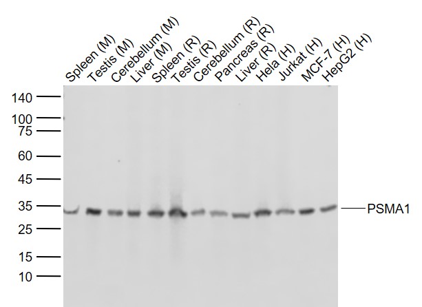
Sample:
Lane 1: Mouse Spleen tissue lysates
Lane 2: Mouse Testis tissue lysates
Lane 3: Mouse Cerebellum tissue lysates
Lane 4: Mouse Liver tissue lysates
Lane 5: Rat Spleen tissue lysates
Lane 6: Rat Testis tissue lysates
Lane 7: Rat Cerebellum tissue lysates
Lane 8: Rat Pancreas tissue lysates
Lane 9: Rat Liver tissue lysates
Lane 10: Human Hela cell lysates
Lane 11: Human Jurkat cell lysates
Lane 12: Human MCF-7 cell lysates
Lane 13: Human HepG2 cell lysates
Primary: Anti-PSMA1 (bsm-54170R) at 1/1000 dilution
Secondary: IRDye800CW Goat Anti-Rabbit IgG at 1/20000 dilution
Predicted band size: 30 kD
Observed band size: 32 kD
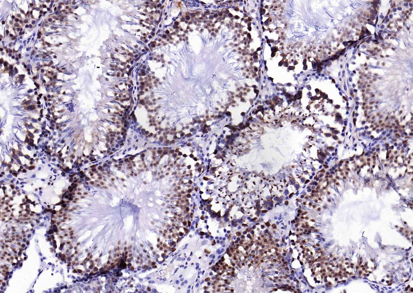
Paraformaldehyde-fixed, paraffin embedded (rat testis); Antigen retrieval by boiling in sodium citrate buffer (pH6.0) for 15min; Block endogenous peroxidase by 3% hydrogen peroxide for 20 minutes; Blocking buffer (normal goat serum) at 37°C for 30min; Antibody incubation with (PSMA1) Monoclonal Antibody, Unconjugated (bsm-54170R) at 1:200 overnight at 4°C, followed by operating according to SP Kit(Rabbit) (sp-0023) instructionsand DAB staining.
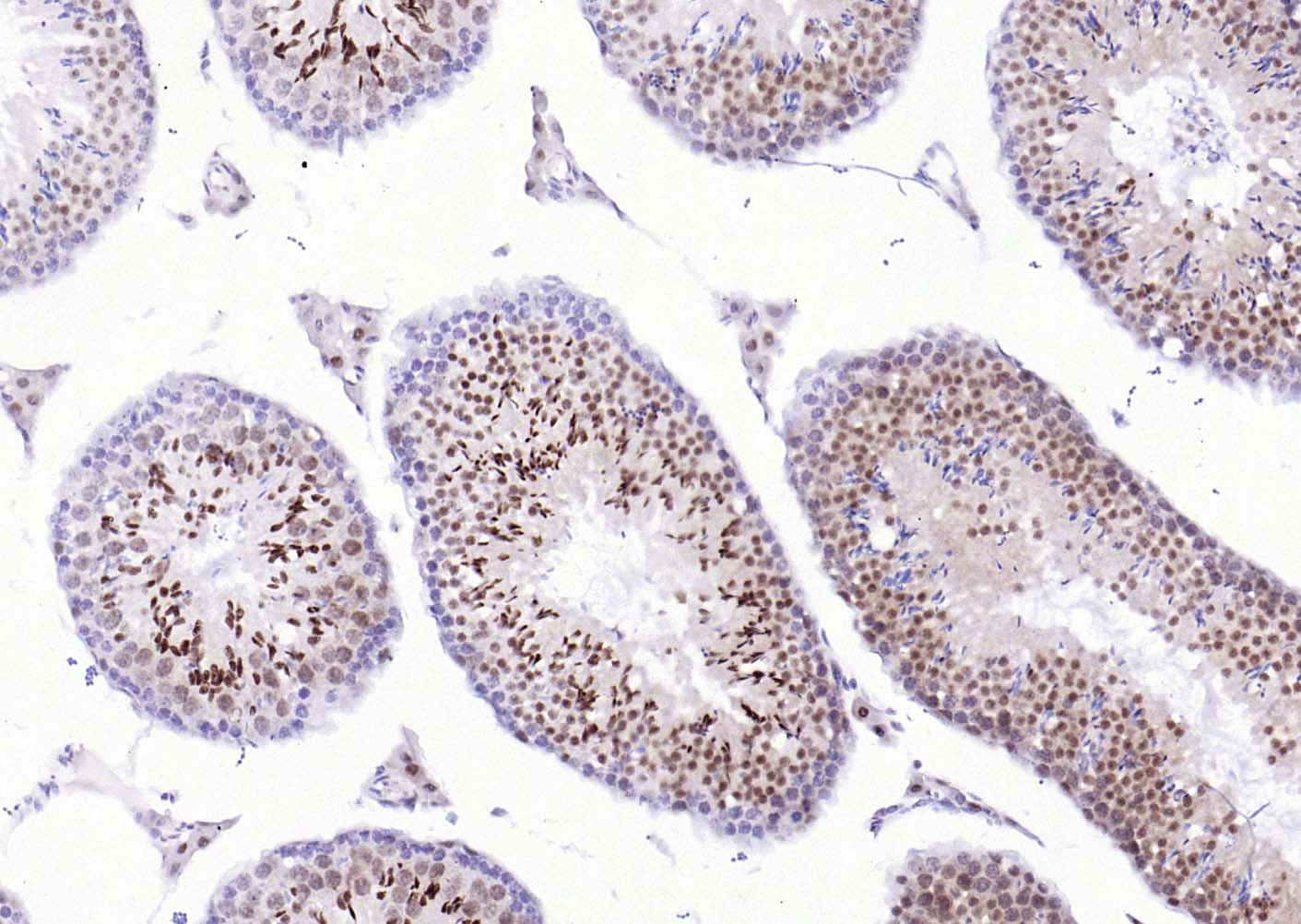
Paraformaldehyde-fixed, paraffin embedded (mouse testis); Antigen retrieval by boiling in sodium citrate buffer (pH6.0) for 15min; Block endogenous peroxidase by 3% hydrogen peroxide for 20 minutes; Blocking buffer (normal goat serum) at 37°C for 30min; Antibody incubation with (PSMA1) Monoclonal Antibody, Unconjugated (bsm-54170R) at 1:200 overnight at 4°C, followed by operating according to SP Kit(Rabbit) (sp-0023) instructionsand DAB staining.
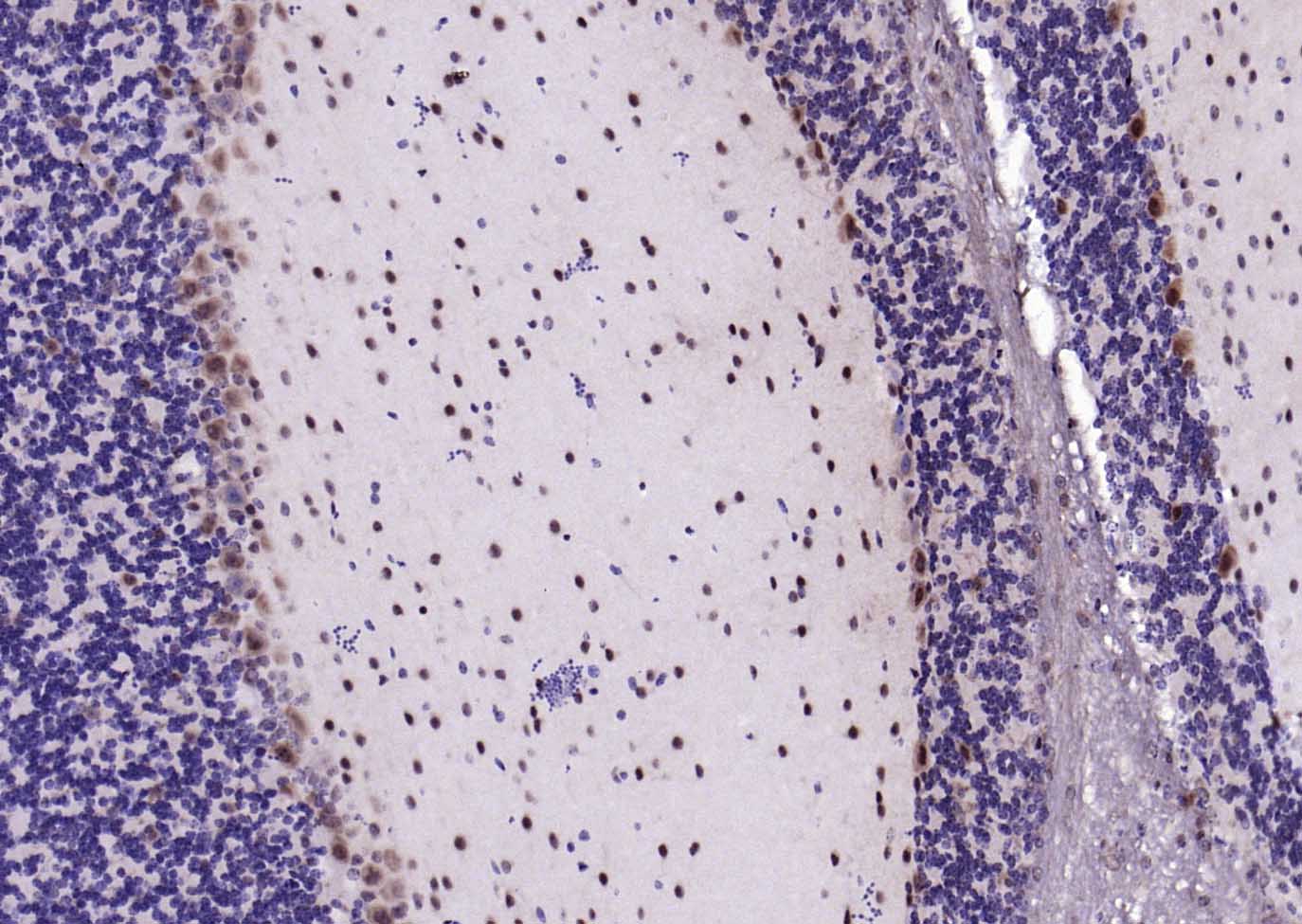
Paraformaldehyde-fixed, paraffin embedded (mouse cerebellum); Antigen retrieval by boiling in sodium citrate buffer (pH6.0) for 15min; Block endogenous peroxidase by 3% hydrogen peroxide for 20 minutes; Blocking buffer (normal goat serum) at 37°C for 30min; Antibody incubation with (PSMA1) Monoclonal Antibody, Unconjugated (bsm-54170R) at 1:200 overnight at 4°C, followed by operating according to SP Kit(Rabbit) (sp-0023) instructionsand DAB staining.
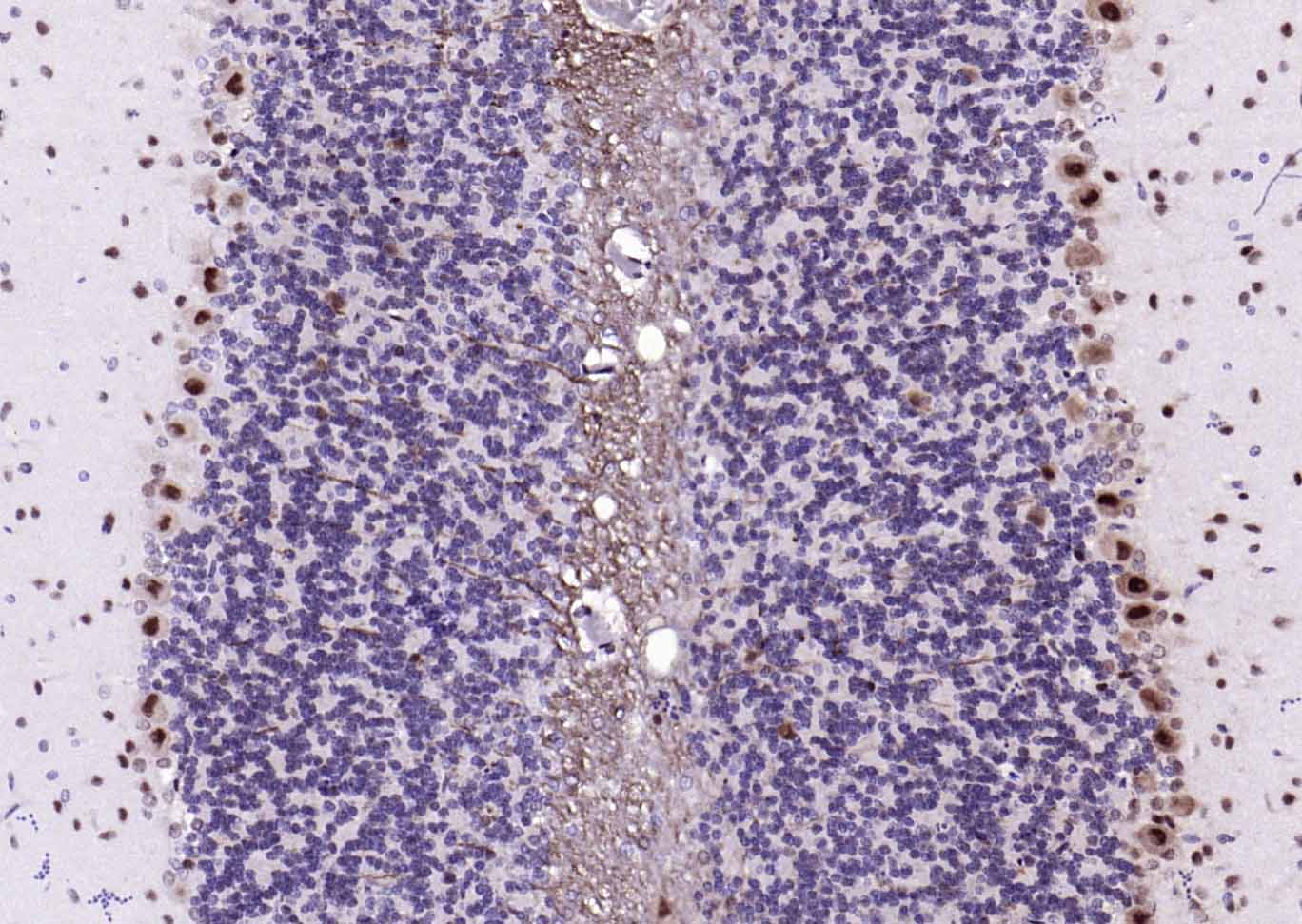
Paraformaldehyde-fixed, paraffin embedded (rat cerebellum); Antigen retrieval by boiling in sodium citrate buffer (pH6.0) for 15min; Block endogenous peroxidase by 3% hydrogen peroxide for 20 minutes; Blocking buffer (normal goat serum) at 37°C for 30min; Antibody incubation with (PSMA1) Monoclonal Antibody, Unconjugated (bsm-54170R) at 1:200 overnight at 4°C, followed by operating according to SP Kit(Rabbit) (sp-0023) instructionsand DAB staining.
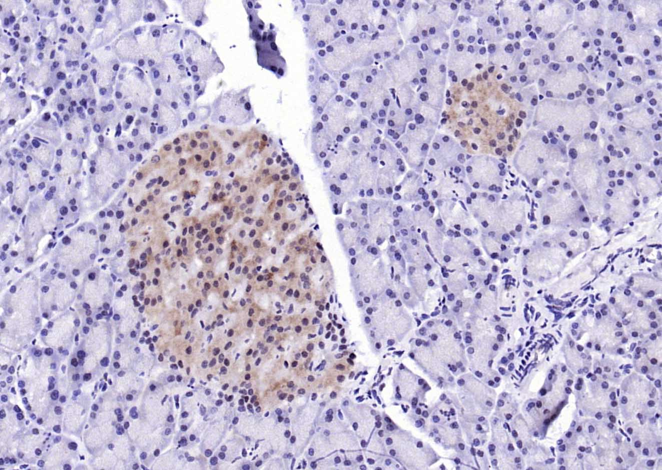
Paraformaldehyde-fixed, paraffin embedded (rat pancreas); Antigen retrieval by boiling in sodium citrate buffer (pH6.0) for 15min; Block endogenous peroxidase by 3% hydrogen peroxide for 20 minutes; Blocking buffer (normal goat serum) at 37°C for 30min; Antibody incubation with (PSMA1) Monoclonal Antibody, Unconjugated (bsm-54170R) at 1:200 overnight at 4°C, followed by operating according to SP Kit(Rabbit) (sp-0023) instructionsand DAB staining.
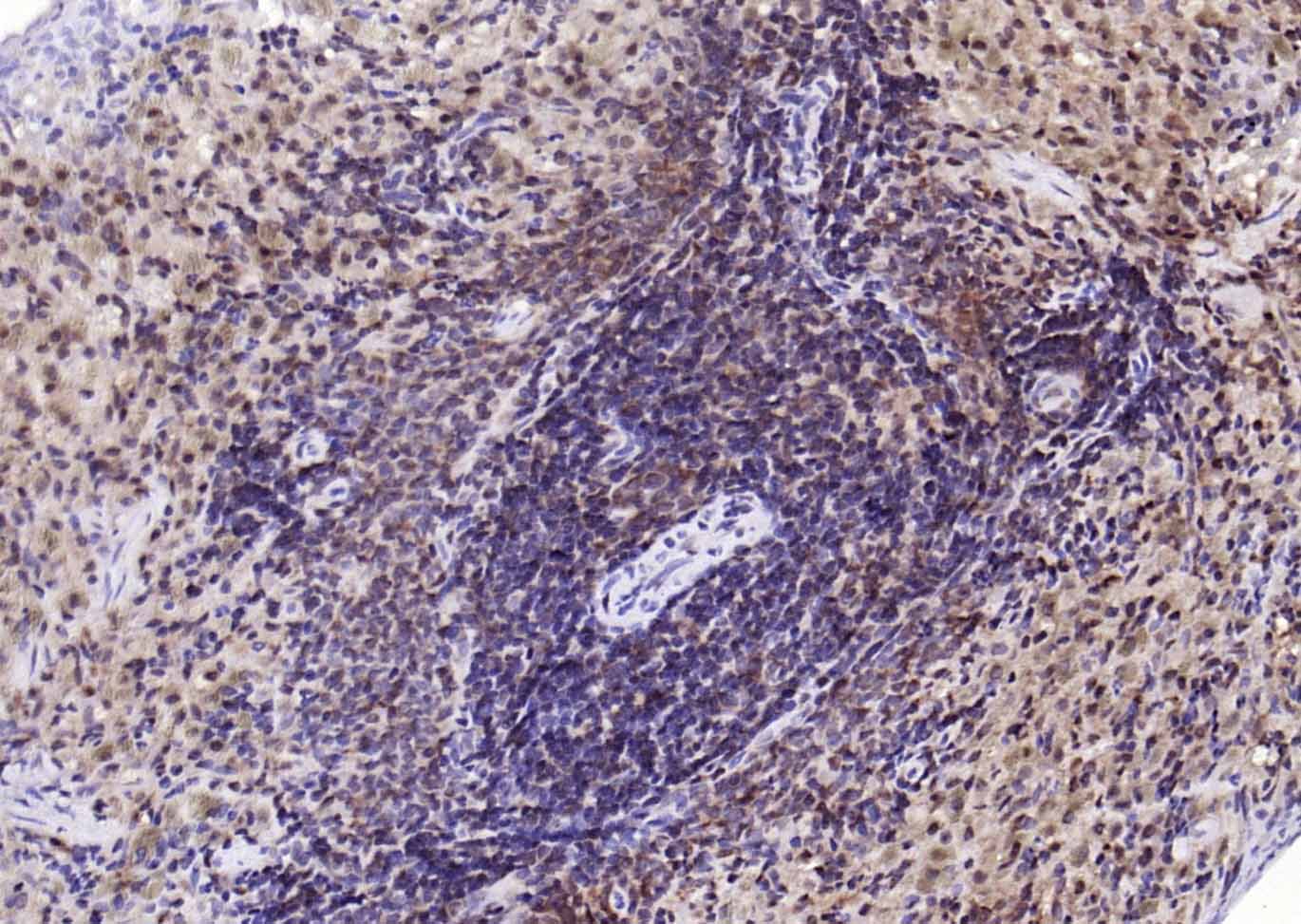
Paraformaldehyde-fixed, paraffin embedded (rat spleen); Antigen retrieval by boiling in sodium citrate buffer (pH6.0) for 15min; Block endogenous peroxidase by 3% hydrogen peroxide for 20 minutes; Blocking buffer (normal goat serum) at 37°C for 30min; Antibody incubation with (PSMA1) Monoclonal Antibody, Unconjugated (bsm-54170R) at 1:200 overnight at 4°C, followed by operating according to SP Kit(Rabbit) (sp-0023) instructionsand DAB staining.
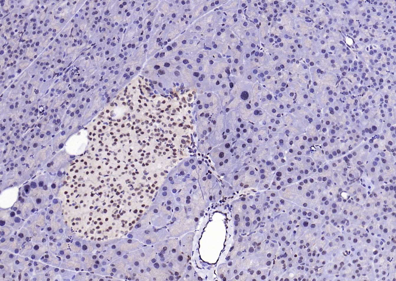
Paraformaldehyde-fixed, paraffin embedded (mouse pancreas); Antigen retrieval by boiling in sodium citrate buffer (pH6.0) for 15min; Block endogenous peroxidase by 3% hydrogen peroxide for 20 minutes; Blocking buffer (normal goat serum) at 37°C for 30min; Antibody incubation with (PSMA1) Monoclonal Antibody, Unconjugated (bsm-54170R) at 1:200 overnight at 4°C, followed by operating according to SP Kit(Rabbit) (sp-0023) instructionsand DAB staining.
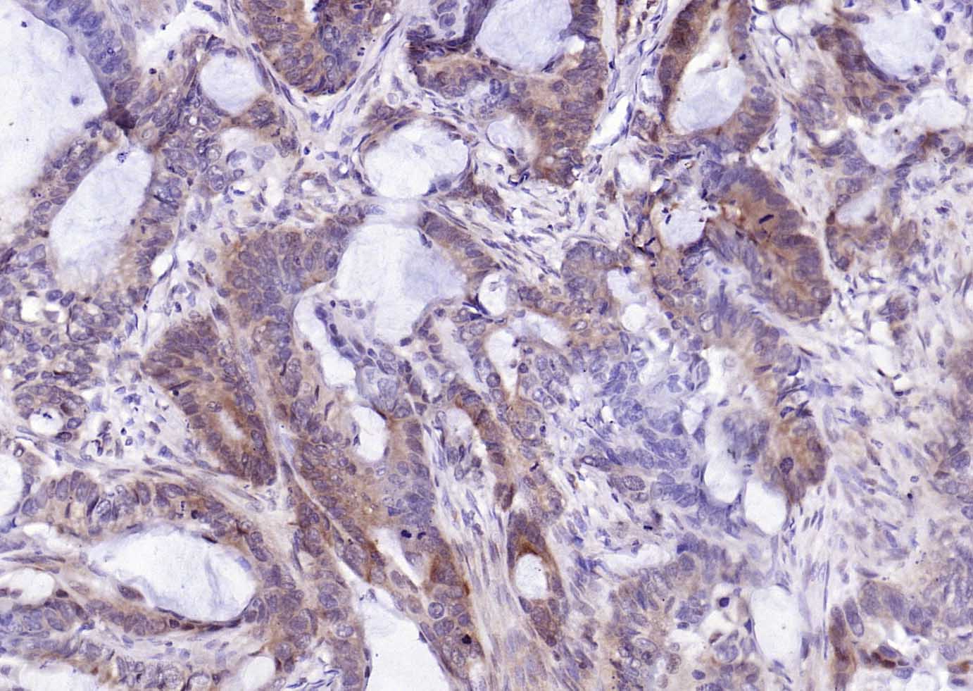
Paraformaldehyde-fixed, paraffin embedded (human colon carcinoma); Antigen retrieval by boiling in sodium citrate buffer (pH6.0) for 15min; Block endogenous peroxidase by 3% hydrogen peroxide for 20 minutes; Blocking buffer (normal goat serum) at 37°C for 30min; Antibody incubation with (PSMA1) Monoclonal Antibody, Unconjugated (bsm-54170R) at 1:200 overnight at 4°C, followed by operating according to SP Kit(Rabbit) (sp-0023) instructionsand DAB staining.
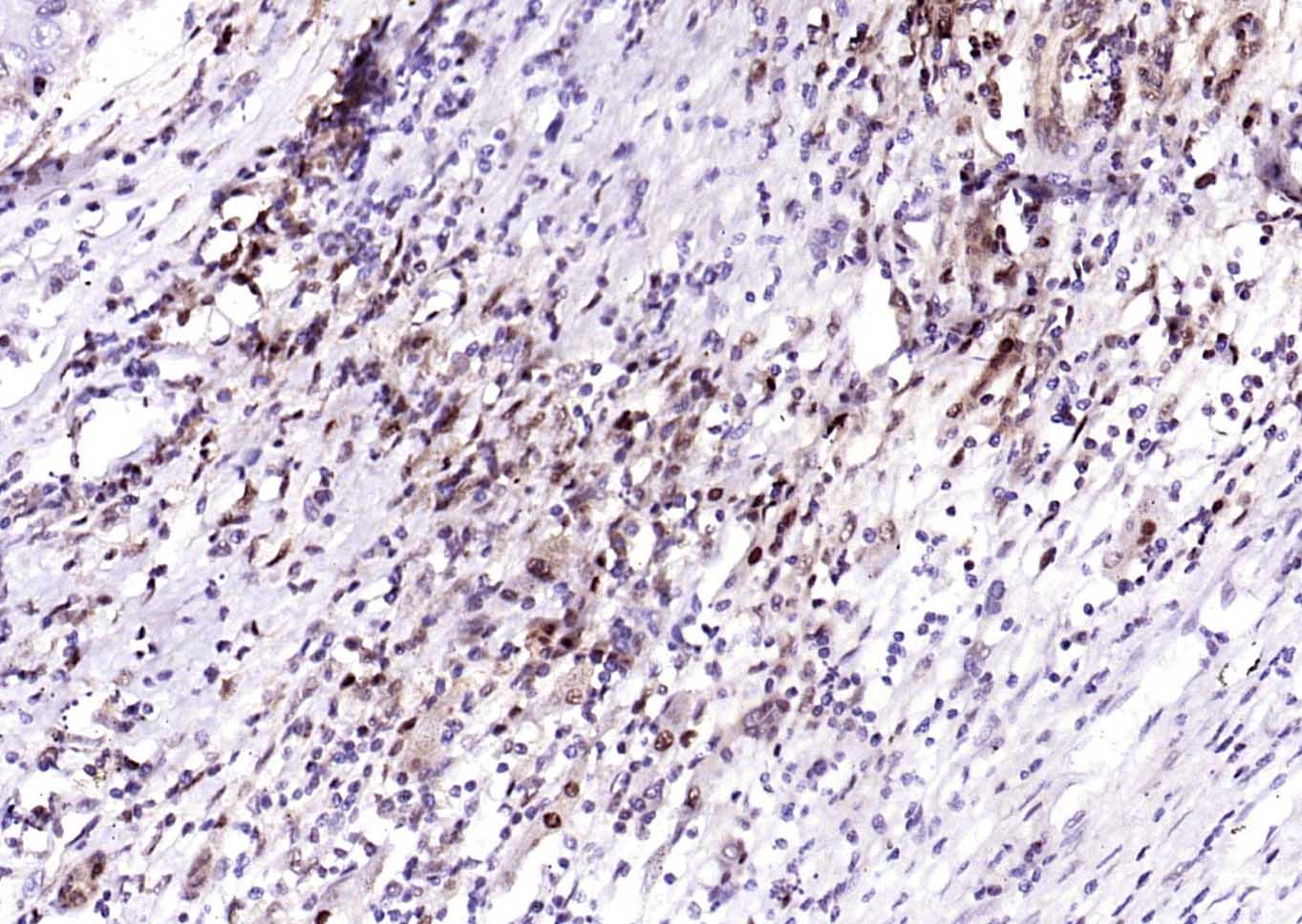
Paraformaldehyde-fixed, paraffin embedded (human liver carcinoma); Antigen retrieval by boiling in sodium citrate buffer (pH6.0) for 15min; Block endogenous peroxidase by 3% hydrogen peroxide for 20 minutes; Blocking buffer (normal goat serum) at 37°C for 30min; Antibody incubation with (PSMA1) Monoclonal Antibody, Unconjugated (bsm-54170R) at 1:200 overnight at 4°C, followed by operating according to SP Kit(Rabbit) (sp-0023) instructionsand DAB staining.
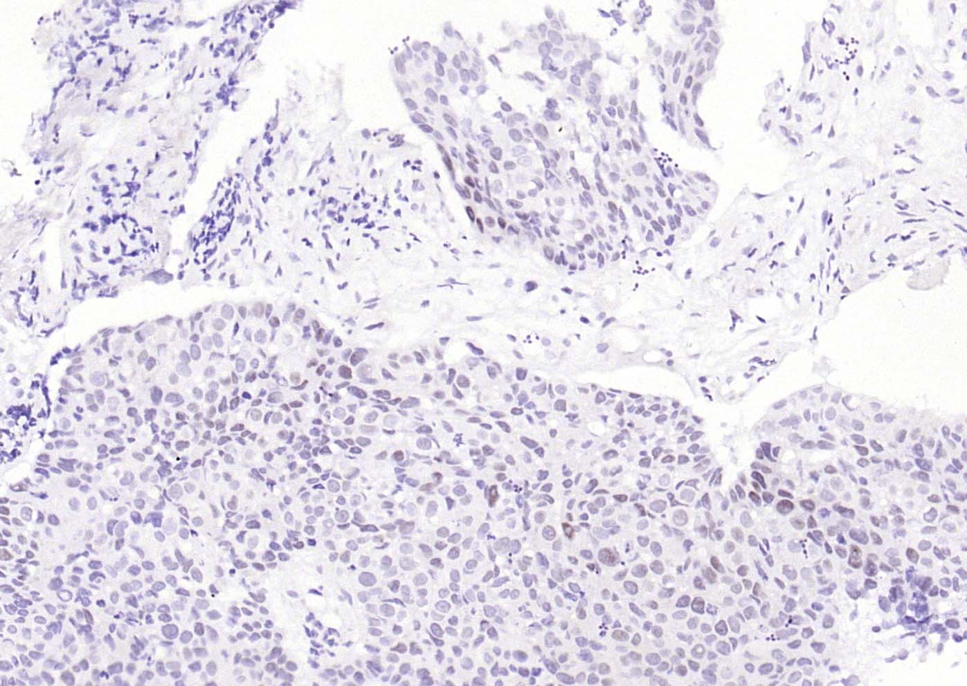
Paraformaldehyde-fixed, paraffin embedded (human breast carcinoma); Antigen retrieval by boiling in sodium citrate buffer (pH6.0) for 15min; Block endogenous peroxidase by 3% hydrogen peroxide for 20 minutes; Blocking buffer (normal goat serum) at 37°C for 30min; Antibody incubation with (PSMA1) Monoclonal Antibody, Unconjugated (bsm-54170R) at 1:200 overnight at 4°C, followed by operating according to SP Kit(Rabbit) (sp-0023) instructionsand DAB staining.
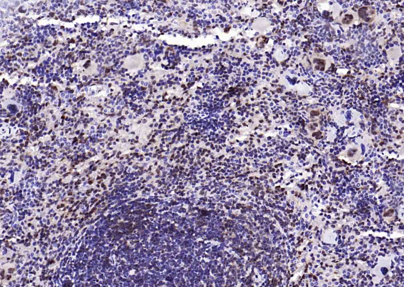
Paraformaldehyde-fixed, paraffin embedded (mouse spleen); Antigen retrieval by boiling in sodium citrate buffer (pH6.0) for 15min; Block endogenous peroxidase by 3% hydrogen peroxide for 20 minutes; Blocking buffer (normal goat serum) at 37°C for 30min; Antibody incubation with (PSMA1) Monoclonal Antibody, Unconjugated (bsm-54170R) at 1:200 overnight at 4°C, followed by operating according to SP Kit(Rabbit) (sp-0023) instructionsand DAB staining.
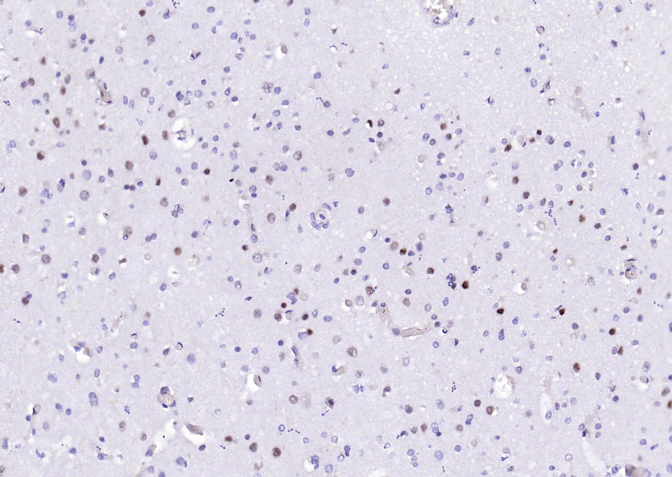
Paraformaldehyde-fixed, paraffin embedded (human brain); Antigen retrieval by boiling in sodium citrate buffer (pH6.0) for 15min; Block endogenous peroxidase by 3% hydrogen peroxide for 20 minutes; Blocking buffer (normal goat serum) at 37°C for 30min; Antibody incubation with (PSMA1) Monoclonal Antibody, Unconjugated (bsm-54170R) at 1:200 overnight at 4°C, followed by operating according to SP Kit(Rabbit) (sp-0023) instructionsand DAB staining.
|
| 1、抗体溶解方法 | |
| 2、抗体修复方式 | |
| 3、常用试剂的配制 | |
| 4、免疫组化操作步骤 | |
| 5、免疫组化问题解答 | |
| 6、Western Blotting 操作步骤 | |
| 7、Western Blotting 问题解答 | |
| 8、关于肽链的设计 | |
| 9、多肽的溶解与保存 | |
| 10、酶标抗体效价测定程序 | |


