
| 产品编号 | bsm-52240R |
| 英文名称 | GLUT1 Recombinant Rabbit mAb |
| 中文名称 | 葡萄糖转运蛋白1重组兔单抗 |
| 别 名 | CSE; DYT17; DYT18; DYT9; EIG12; GLUT; GLUT-1; GLUT1; GLUT1DS; HTLVR; PED; SDCHCN; GT1; M100200; Rgsc200; GLUTB; GTG1; Gtg3; RATGTG1; GTR1_HUMAN; SLC2A1; Glucose transporter type 1, erythrocyte/brain (GLUT-1); HepG2 glucose transporter; GTR1_MOUSE; Glucose |
 |
Specific References (1) | bsm-52240R has been referenced in 1 publications.
[IF=15.153] Shuaijun Lu. et al. Nanoengineering a Zeolitic Imidazolate Framework-8 Capable of Manipulating Energy Metabolism against Cancer Chemo-Phototherapy Resistance. SMALL. 2022 Oct;:2204926 WB ; Mouse, Human.
|
| 研究领域 | 肿瘤 免疫学 生长因子和激素 转运蛋白 |
| 抗体来源 | Rabbit |
| 克隆类型 | Recombinant |
| 克 隆 号 | 2D5 |
| 交叉反应 | Human,Mouse,Rat |
| 产品应用 | IHC-P=1:200-1000,IHC-F=1:200-1000,IF=1:200-1000,ICC/IF=1:50-200
not yet tested in other applications. optimal dilutions/concentrations should be determined by the end user. |
| 理论分子量 | 54 kDa |
| 细胞定位 | 细胞膜 细胞外基质 |
| 性 状 | Liquid |
| 浓 度 | 1mg/ml |
| 免 疫 原 | A synthesized peptide derived from human GLUT1: 450-492 |
| 亚 型 | IgG |
| 纯化方法 | affinity purified by Protein A |
| 缓 冲 液 | 0.01M TBS (pH7.4) with 1% BSA, 0.02% Proclin300 and 50% Glycerol. |
| 保存条件 | Shipped at 4℃. Store at -20℃ for one year. Avoid repeated freeze/thaw cycles. |
| 注意事项 | This product as supplied is intended for research use only, not for use in human, therapeutic or diagnostic applications. |
| PubMed | PubMed |
| 产品介绍 |
This gene encodes a major glucose transporter in the mammalian blood-brain barrier. Mutations in this gene have been found in a family with paroxysmal exertion-induced dyskinesia. [provided by RefSeq, Jul 2008]. Function: Facilitative glucose transporter. This isoform may be responsible for constitutive or basal glucose uptake. Has a very broad substrate specificity; can transport a wide range of aldoses including both pentoses and hexoses. Subcellular Location: Cell membrane; Multi-pass membrane protein. Melanosome. Note=Localizes primarily at the cell surface. Identified by mass spectrometry in melanosome fractions from stage I to stage IV. Tissue Specificity: Expressed at variable levels in many human tissues. Post-translational modifications: Phosphorylated upon DNA damage, probably by ATM or ATR. DISEASE: Defects in SLC2A1 are the cause of GLUT1 deficiency syndrome type 1 (GLUT1DS1) [MIM:606777]; also known as blood-brain barrier glucose transport defect. A neurologic disorder showing wide phenotypic variability. The most severe 'classic' phenotype comprises infantile-onset epileptic encephalopathy associated with delayed development, acquired microcephaly, motor incoordination, and spasticity. Onset of seizures, usually characterized by apneic episodes, staring spells, and episodic eye movements, occurs within the first 4 months of life. Other paroxysmal findings include intermittent ataxia, confusion, lethargy, sleep disturbance, and headache. Varying degrees of cognitive impairment can occur, ranging from learning disabilities to severe mental retardation. Defects in SLC2A1 are the cause of GLUT1 deficiency syndrome type 2 (GLUT1DS2) [MIM:612126]. A clinically variable disorder characterized primarily by onset in childhood of paroxysmal exercise-induced dyskinesia. The dyskinesia involves transient abnormal involuntary movements, such as dystonia and choreoathetosis, induced by exercise or exertion, and affecting the exercised limbs. Some patients may also have epilepsy, most commonly childhood absence epilepsy. Mild mental retardation may also occur. In some patients involuntary exertion-induced dystonic, choreoathetotic, and ballistic movements may be associated with macrocytic hemolytic anemia. Similarity: Belongs to the major facilitator superfamily. Sugar transporter (TC 2.A.1.1) family. Glucose transporter subfamily. SWISS: P11166 Gene ID: 6513 Database links: Entrez Gene: 6513 Human Entrez Gene: 20525 Mouse Omim: 138140 Human SwissProt: P11166 Human SwissProt: P17809 Mouse Unigene: 473721 Human Unigene: 721551 Human Unigene: 21002 Mouse Unigene: 3205 Rat |
| 产品图片 |
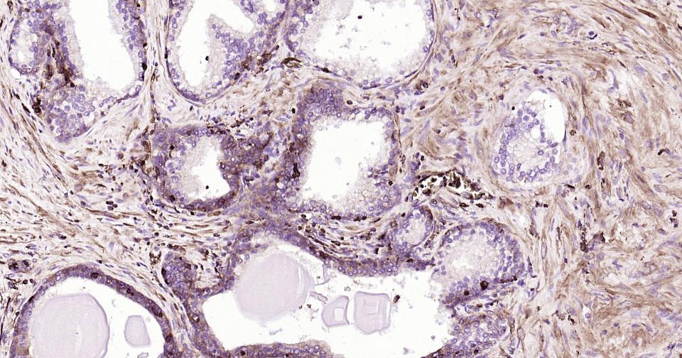
Paraformaldehyde-fixed, paraffin embedded Human Prostate; Antigen retrieval by boiling in sodium citrate buffer (pH6.0) for 15 min; Antibody incubation with GLUT1 Monoclonal Antibody, Unconjugated (bsm-52240R) at 1:200 overnight at 4°C, followed by conjugation to the bs-0295G-HRP and DAB (C-0010) staining.
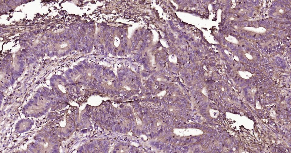
Paraformaldehyde-fixed, paraffin embedded Human Colon Cancer; Antigen retrieval by boiling in sodium citrate buffer (pH6.0) for 15 min; Antibody incubation with GLUT1 Monoclonal Antibody, Unconjugated (bsm-52240R) at 1:200 overnight at 4°C, followed by conjugation to the bs-0295G-HRP and DAB (C-0010) staining.
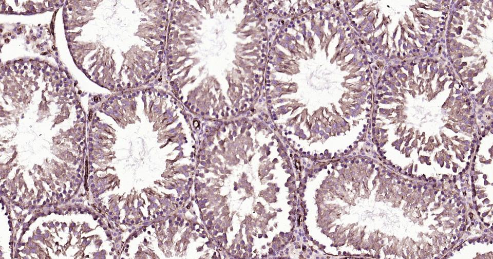
Paraformaldehyde-fixed, paraffin embedded Rat Testicles; Antigen retrieval by boiling in sodium citrate buffer (pH6.0) for 15 min; Antibody incubation with GLUT1 Monoclonal Antibody, Unconjugated (bsm-52240R) at 1:200 overnight at 4°C, followed by conjugation to the bs-0295G-HRP and DAB (C-0010) staining.
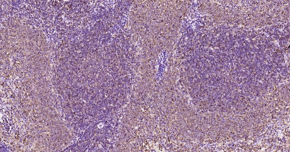
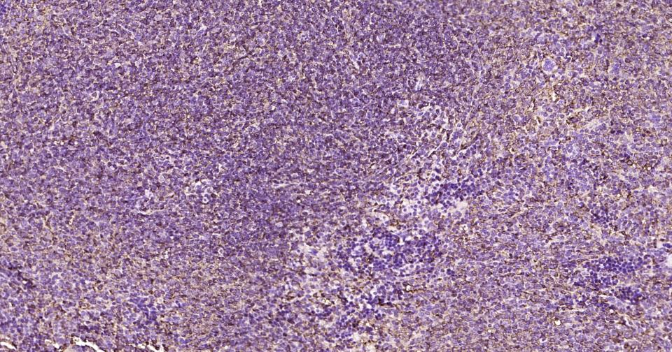
Paraformaldehyde-fixed, paraffin embedded Rat Spleen; Antigen retrieval by boiling in sodium citrate buffer (pH6.0) for 15 min; Antibody incubation with GLUT1 Monoclonal Antibody, Unconjugated (bsm-52240R) at 1:200 overnight at 4°C, followed by conjugation to the bs-0295G-HRP and DAB (C-0010) staining.
Paraformaldehyde-fixed, paraffin embedded Mouse Spleen; Antigen retrieval by boiling in sodium citrate buffer (pH6.0) for 15 min; Antibody incubation with GLUT1 Monoclonal Antibody, Unconjugated (bsm-52240R) at 1:200 overnight at 4°C, followed by conjugation to the bs-0295G-HRP and DAB (C-0010) staining.
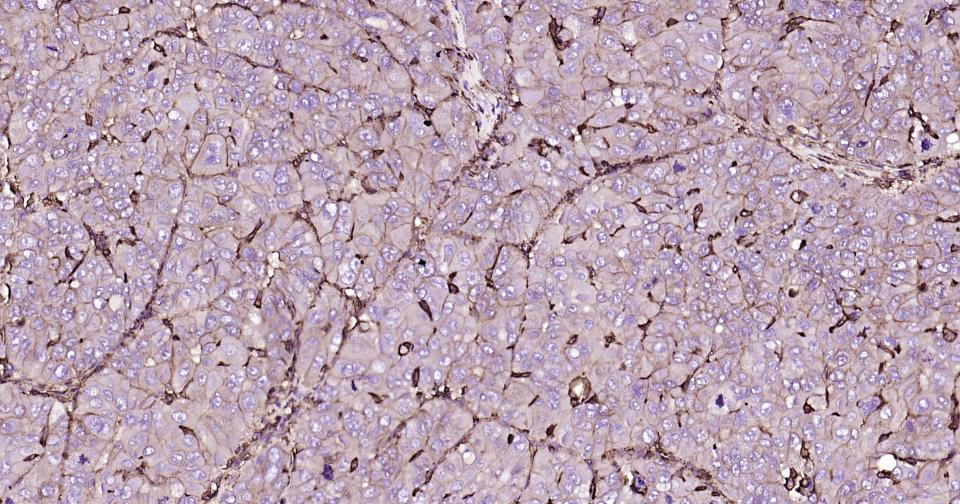
Paraformaldehyde-fixed, paraffin embedded Human Liver Caner; Antigen retrieval by boiling in sodium citrate buffer (pH6.0) for 15 min; Antibody incubation with GLUT1 Monoclonal Antibody, Unconjugated (bsm-52240R) at 1:200 overnight at 4°C, followed by conjugation to the bs-0295G-HRP and DAB (C-0010) staining.

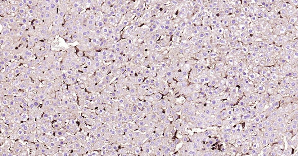
Paraformaldehyde-fixed, paraffin embedded Human Liver; Antigen retrieval by boiling in sodium citrate buffer (pH6.0) for 15 min; Antibody incubation with GLUT1 Monoclonal Antibody, Unconjugated (bsm-52240R) at 1:200 overnight at 4°C, followed by conjugation to the bs-0295G-HRP and DAB (C-0010) staining.
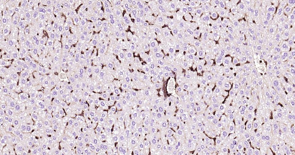
Paraformaldehyde-fixed, paraffin embedded Rat Liver; Antigen retrieval by boiling in sodium citrate buffer (pH6.0) for 15 min; Antibody incubation with GLUT1 Monoclonal Antibody, Unconjugated (bsm-52240R) at 1:200 overnight at 4°C, followed by conjugation to the bs-0295G-HRP and DAB (C-0010) staining.
Paraformaldehyde-fixed, paraffin embedded Mouse Liver; Antigen retrieval by boiling in sodium citrate buffer (pH6.0) for 15 min; Antibody incubation with GLUT1 Monoclonal Antibody, Unconjugated (bsm-52240R) at 1:200 overnight at 4°C, followed by conjugation to the bs-0295G-HRP and DAB (C-0010) staining.
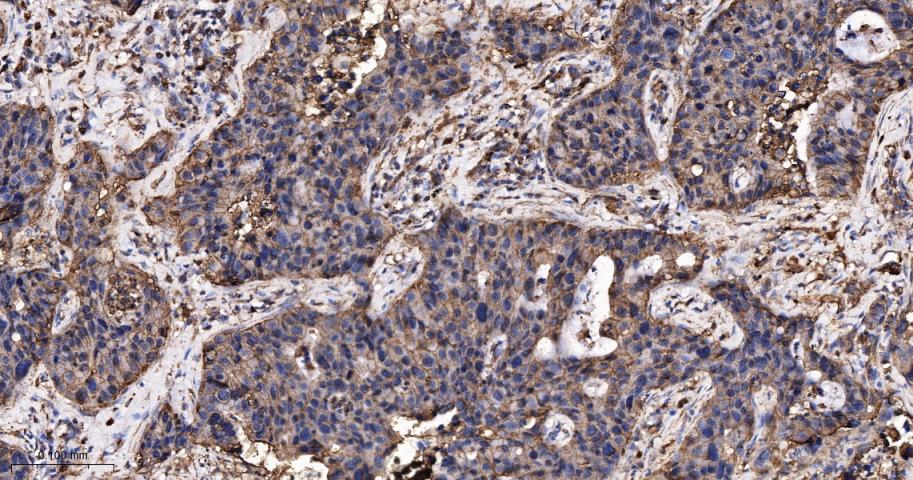
Paraformaldehyde-fixed, paraffin embedded Human Lung Cancer; Antigen retrieval by boiling in sodium citrate buffer (pH6.0) for 15 min; Antibody incubation with GLUT1 Monoclonal Antibody, Unconjugated (bsm-52240R) at 1:500 overnight at 4°C, followed by conjugation to the bs-0295G-HRP and DAB (C-0010) staining.
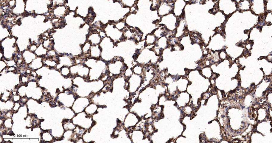
Paraformaldehyde-fixed, paraffin embedded Rat Lung; Antigen retrieval by boiling in sodium citrate buffer (pH6.0) for 15 min; Antibody incubation with GLUT1 Monoclonal Antibody, Unconjugated (bsm-52240R) at 1:500 overnight at 4°C, followed by conjugation to the bs-0295G-HRP and DAB (C-0010) staining.
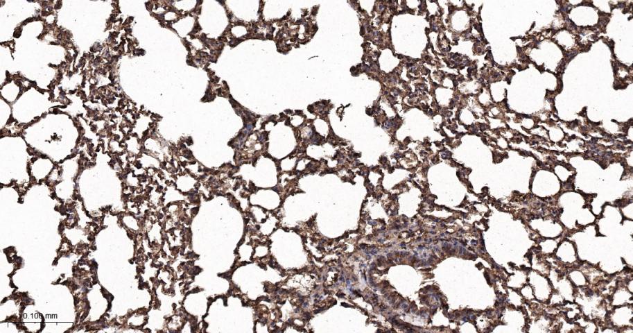
Paraformaldehyde-fixed, paraffin embedded Mouse Lung; Antigen retrieval by boiling in sodium citrate buffer (pH6.0) for 15 min; Antibody incubation with GLUT1 Monoclonal Antibody, Unconjugated (bsm-52240R) at 1:500 overnight at 4°C, followed by conjugation to the bs-0295G-HRP and DAB (C-0010) staining.
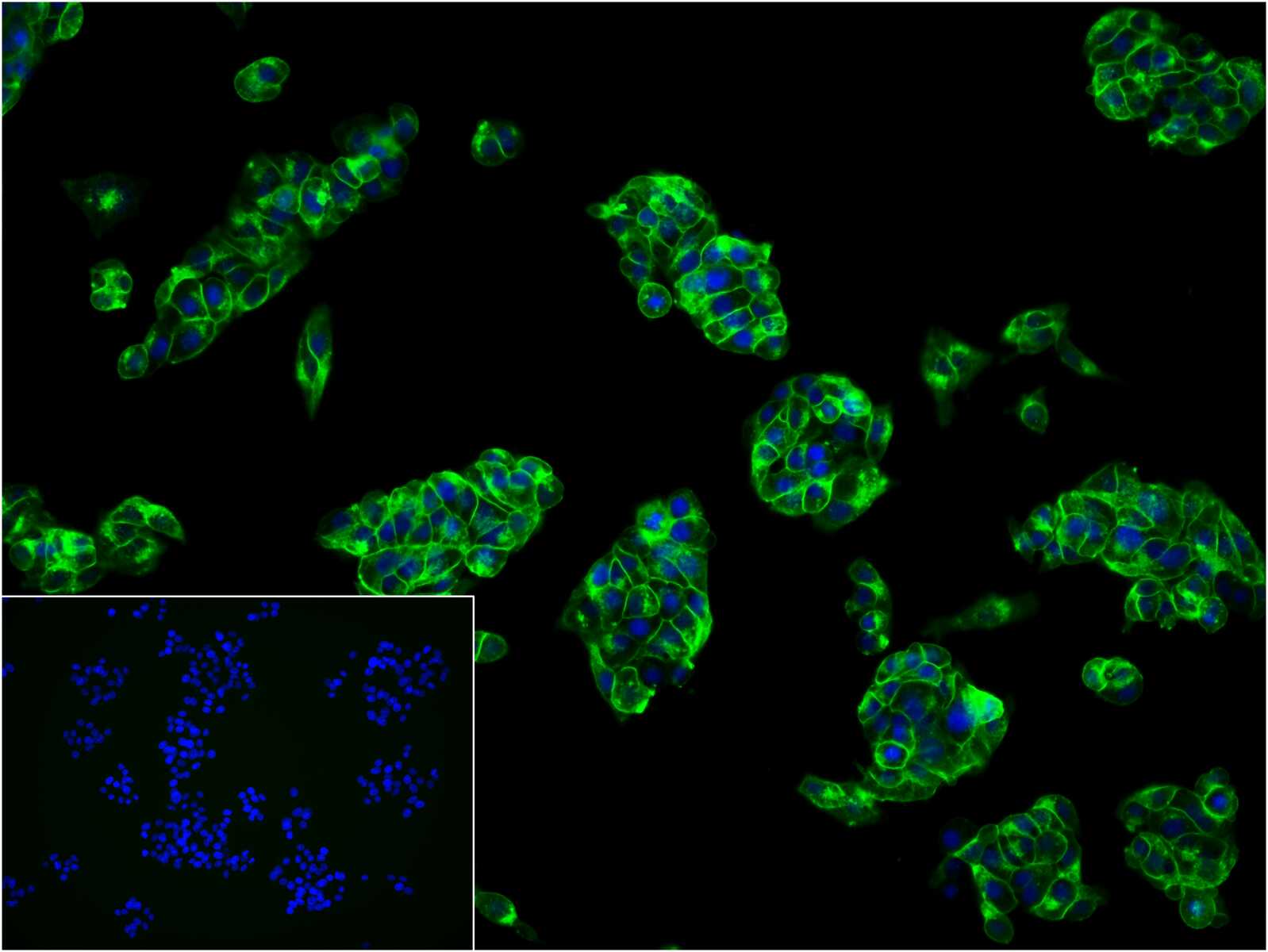
4% Paraformaldehyde-fixed HepG2 (H) cell; Triton X-100 at r.t. for 20 min; Antibody incubation with (GLUT1) monoclonal Antibody, unconjugated (bsm-52240R) 1:100, 90 min at 37°C; followed by BF488 conjugated Goat Anti-Rabbit IgG antibody (green, bs-60295G-BF488) at 37°C for 90 min, DAPI (blue, C02-04002) was used to stain the cell nuclei. PBS instead of the primary antibody was used as the blank control.
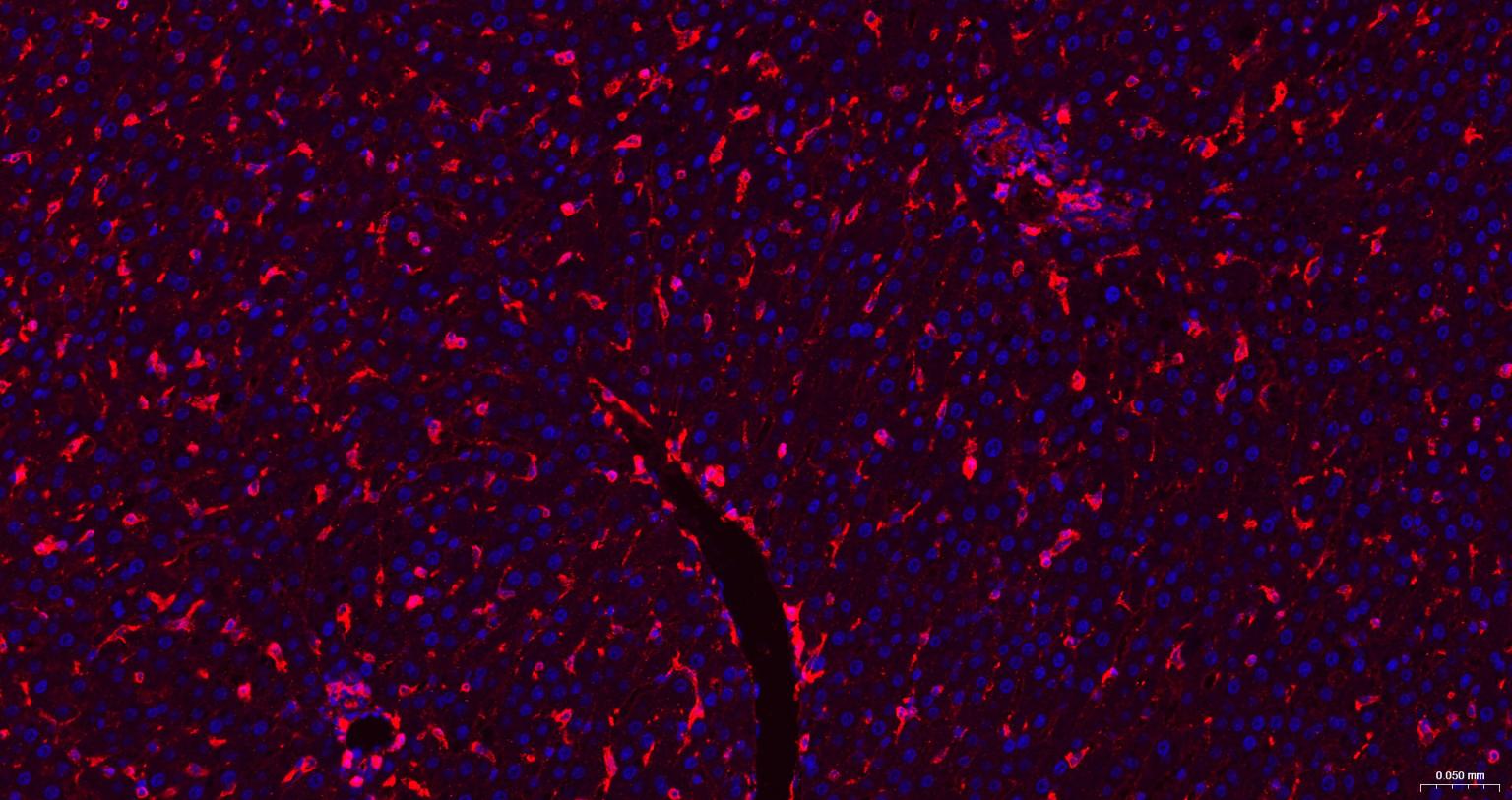
Paraformaldehyde-fixed, paraffin embedded Rat Liver; Antigen retrieval by boiling in sodium citrate buffer (pH6.0) for 15 min; The section was incubated with GLUT1 Monoclonal Antibody, Unconjugated (bsm-52240R) at 1:200 overnight at 4°C. Followed by conjugated Goat Anti-Rabbit IgG antibody (Red, bs-0295G-BF594), DAPI (blue, C02-04002) was used to stain the cell nuclei.
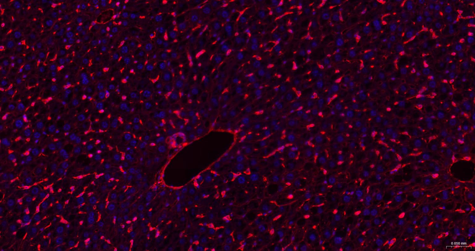
Paraformaldehyde-fixed, paraffin embedded Mouse Liver; Antigen retrieval by boiling in sodium citrate buffer (pH6.0) for 15 min; The section was incubated with GLUT1 Monoclonal Antibody, Unconjugated (bsm-52240R) at 1:200 overnight at 4°C. Followed by conjugated Goat Anti-Rabbit IgG antibody (Red, bs-0295G-BF594), DAPI (blue, C02-04002) was used to stain the cell nuclei.
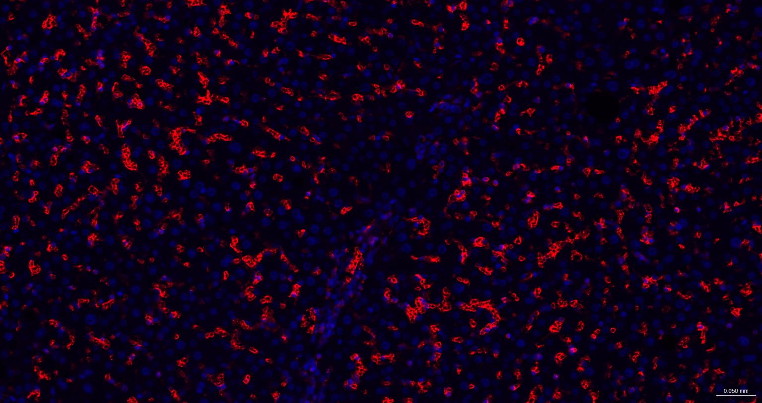
Paraformaldehyde-fixed, paraffin embedded Human Liver; Antigen retrieval by boiling in sodium citrate buffer (pH6.0) for 15 min; The section was incubated with GLUT1 Monoclonal Antibody, Unconjugated (bsm-52240R) at 1:200 overnight at 4°C. Followed by conjugated Goat Anti-Rabbit IgG antibody (Red, bs-0295G-BF594), DAPI (blue, C02-04002) was used to stain the cell nuclei.
|
| 1、抗体溶解方法 | |
| 2、抗体修复方式 | |
| 3、常用试剂的配制 | |
| 4、免疫组化操作步骤 | |
| 5、免疫组化问题解答 | |
| 6、Western Blotting 操作步骤 | |
| 7、Western Blotting 问题解答 | |
| 8、关于肽链的设计 | |
| 9、多肽的溶解与保存 | |
| 10、酶标抗体效价测定程序 | |


