
| 产品编号 | bsm-52938R |
| 英文名称 | TNFR2 Recombinant Rabbit mAb |
| 中文名称 | 肿瘤坏死因子受体2重组兔单抗 |
| 别 名 | CD120b; TBPII; TNF-R-II; TNF-R75; TNFBR; TNFR1B; TNFR2; TNFR80; p75; p75TNFR; TNF-R2; TNF-alphaR2; TNFRII; TNFalpha-R2; Tnfr-1; TNR1B_HUMAN; TNFRSF1B; Tumor necrosis factor receptor 2 (TNF-R2); Tumor necrosis factor receptor type II (TNF-RII | TNFR-II); p |
| 研究领域 | 肿瘤 细胞生物 神经生物学 信号转导 细胞凋亡 细胞膜受体 |
| 抗体来源 | Rabbit |
| 克隆类型 | Recombinant |
| 克 隆 号 | 7D3 |
| 交叉反应 | Human (predicted: Mouse,Rat) |
| 产品应用 | IHC-P=1:50-200,IHC-F=1:50-200,IF=1:50-200
not yet tested in other applications. optimal dilutions/concentrations should be determined by the end user. |
| 理论分子量 | 46 kDa |
| 细胞定位 | 细胞膜 分泌型蛋白 |
| 性 状 | Liquid |
| 浓 度 | 1mg/ml |
| 免 疫 原 | A synthesized peptide derived from human TNFRSF1B: 415-461/461 |
| 亚 型 | IgG |
| 纯化方法 | affinity purified by Protein A |
| 缓 冲 液 | 0.01M TBS (pH7.4) with 1% BSA, 0.02% Proclin300 and 50% Glycerol. |
| 保存条件 | Shipped at 4℃. Store at -20℃ for one year. Avoid repeated freeze/thaw cycles. |
| 注意事项 | This product as supplied is intended for research use only, not for use in human, therapeutic or diagnostic applications. |
| PubMed | PubMed |
| 产品介绍 |
The protein encoded by this gene is a member of the TNF-receptor superfamily. This protein and TNF-receptor 1 form a heterocomplex that mediates the recruitment of two anti-apoptotic proteins, c-IAP1 and c-IAP2, which possess E3 ubiquitin ligase activity. The function of IAPs in TNF-receptor signalling is unknown, however, c-IAP1 is thought to potentiate TNF-induced apoptosis by the ubiquitination and degradation of TNF-receptor-associated factor 2, which mediates anti-apoptotic signals. Knockout studies in mice also suggest a role of this protein in protecting neurons from apoptosis by stimulating antioxidative pathways. [provided by RefSeq, Jul 2008] Function: Receptor with high affinity for TNFSF2/TNF-alpha and approximately 5-fold lower affinity for homotrimeric TNFSF1/lymphotoxin-alpha. The TRAF1/TRAF2 complex recruits the apoptotic suppressors BIRC2 and BIRC3 to TNFRSF1B/TNFR2. This receptor mediates most of the metabolic effects of TNF-alpha. Isoform 2 blocks TNF-alpha-induced apoptosis, which suggests that it regulates TNF-alpha function by antagonizing its biological activity. Subcellular Location: Secreted and Cell membrane. Post-translational modifications: Phosphorylated; mainly on serine residues and with a very low level on threonine residues. A soluble form (tumor necrosis factor binding protein 2) is produced from the membrane form by proteolytic processing. Similarity: Contains 4 TNFR-Cys repeats. SWISS: P20333 Gene ID: 7133 Database links: Entrez Gene: 7133 Human Entrez Gene: 21938 Mouse Omim: 191191 Human SwissProt: P20333 Human SwissProt: P25119 Mouse Unigene: 256278 Human Unigene: 235328 Mouse Unigene: 83633 Rat |
| 产品图片 |
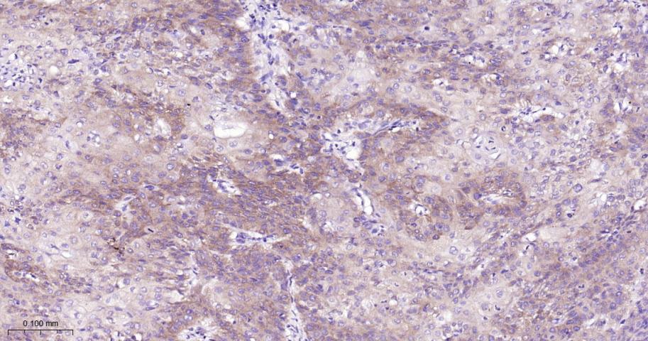
Paraformaldehyde-fixed, paraffin embedded Human Kidney Cancer; Antigen retrieval by boiling in sodium citrate buffer (pH6.0) for 15 min; The section was incubated with TNFR2 Monoclonal Antibody, Unconjugated (bsm-52938R) at 1:200 overnight at 4°C, followed by conjugation to the bs-0295G-HRP and DAB (C-0010) staining.
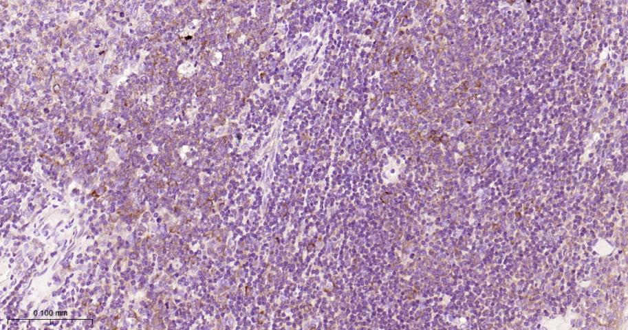
Paraformaldehyde-fixed, paraffin embedded Human Chronic tonsillitis; Antigen retrieval by boiling in sodium citrate buffer (pH6.0) for 15 min; The section was incubated with TNFR2 Monoclonal Antibody, Unconjugated (bsm-52938R) at 1:200 overnight at 4°C, followed by conjugation to the bs-0295G-HRP and DAB (C-0010) staining.
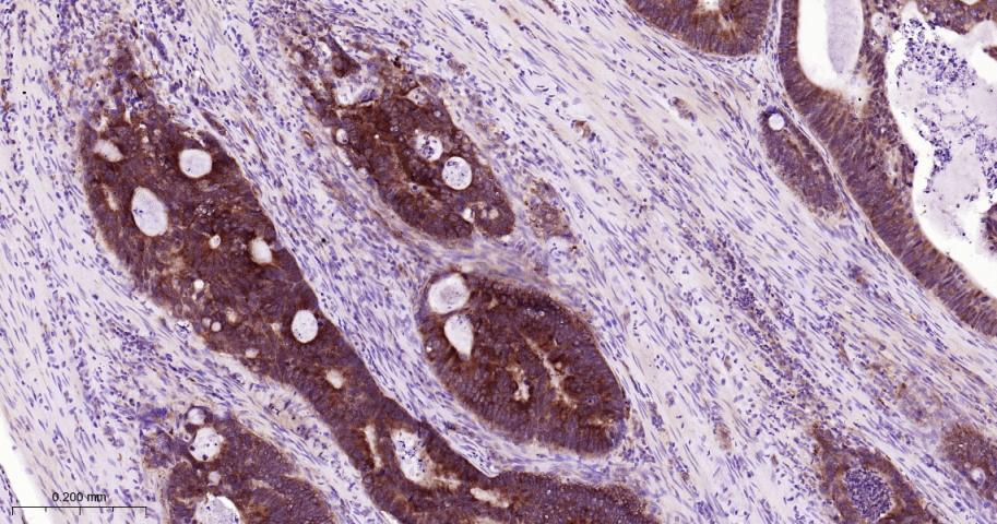
Paraformaldehyde-fixed, paraffin embedded Human Colon Cancer; Antigen retrieval by boiling in sodium citrate buffer (pH6.0) for 15 min; The section was incubated with TNFR2 Monoclonal Antibody, Unconjugated (bsm-52938R) at 1:200 overnight at 4°C, followed by conjugation to the bs-0295G-HRP and DAB (C-0010) staining.
|
| 1、抗体溶解方法 | |
| 2、抗体修复方式 | |
| 3、常用试剂的配制 | |
| 4、免疫组化操作步骤 | |
| 5、免疫组化问题解答 | |
| 6、Western Blotting 操作步骤 | |
| 7、Western Blotting 问题解答 | |
| 8、关于肽链的设计 | |
| 9、多肽的溶解与保存 | |
| 10、酶标抗体效价测定程序 | |


