
| 产品编号 | bsm-54132R |
| 英文名称 | AIF1 Recombinant Rabbit mAb |
| 中文名称 | 离子钙接头蛋白单克隆抗体 |
| 别 名 | AIF-1; IBA1; IRT-1; IRT1; D17H6S50E; G1; BART-1; Bart1; mrf-1; AIF1_HUMAN; AIF1; Ionized calcium-binding adapter molecule 1; Protein G1; AIF1_MOUSE; AIF1_RAT; Microglia response factor; Mrf1; |
| 研究领域 | 细胞生物 免疫学 神经生物学 |
| 抗体来源 | Rabbit |
| 克隆类型 | Recombinant |
| 克 隆 号 | 9A3 |
| 交叉反应 | Human,Mouse,Rat |
| 产品应用 | WB=1:1000-2000,IHC-P=1:200-1000,IHC-F=1:200-1000,IF=1:200-1000
not yet tested in other applications. optimal dilutions/concentrations should be determined by the end user. |
| 理论分子量 | 17 kDa |
| 检测分子量 | 16 |
| 细胞定位 | 细胞浆 细胞膜 |
| 性 状 | Liquid |
| 浓 度 | 1mg/ml |
| 免 疫 原 | A synthesized peptide derived from human IBA1: 1-38 |
| 亚 型 | IgG |
| 纯化方法 | affinity purified by Protein A |
| 缓 冲 液 | 0.01M TBS (pH7.4) with 1% BSA, 0.02% Proclin300 and 50% Glycerol. |
| 保存条件 | Shipped at 4℃. Store at -20℃ for one year. Avoid repeated freeze/thaw cycles. |
| 注意事项 | This product as supplied is intended for research use only, not for use in human, therapeutic or diagnostic applications. |
| PubMed | PubMed |
| 产品介绍 |
Allograft Inflammatory Factor-1 (AIF1)or ionized calcium-binding adaptor molecule 1 (Iba1) is expressed selectively in microglia/macrophages and is a Ca2+-binding peptide produced by activated monocytes and microglial cells. It has been suggested that AIF1 expression is associated with chronic inflammatory processes. AIF1 is expressed by activated monocytes and might participate in a variety of pathogenic processes in the mammalian brain and in chronic transplant rejection. It has been shown to be expressed early and persistently in chronically rejecting cardiac allografts but not in cardiac syngrafts and host hearts. Function: Actin-binding protein that enhances membrane ruffling and RAC activation. Enhances the actin-bundling activity of LCP1. Binds calcium. Plays a role in RAC signaling and in phagocytosis. May play a role in macrophage activation and function. Promotes the proliferation of vascular smooth muscle cells and of T-lymphocytes. Enhances lymphocyte migration. Plays a role in vascular inflammation. Subunit: Homodimer (Potential). Monomer. Interacts with LCP1. Subcellular Location: Cytoplasm, cytoskeleton. Cell projection, ruffle membrane; Peripheral membrane protein; Cytoplasmic side. Note=Associated with the actin cytoskeleton at membrane ruffles and at sites of phagocytosis. Tissue Specificity: Detected in T-lymphocytes and peripheral blood mononuclear cells. Similarity: Contains 2 EF-hand domains. SWISS: P55008 Gene ID: 199 |
| 产品图片 |
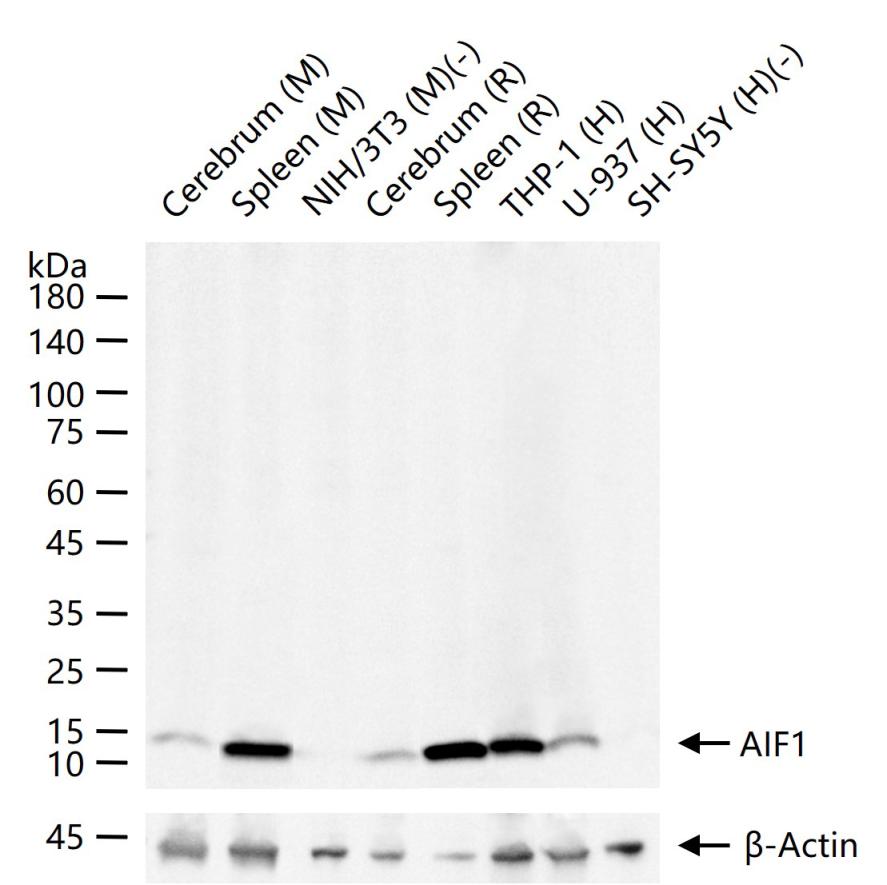
25 ug total protein per lane of various lysates (see on figure) probed with AIF1 (9A3) monoclonal antibody, unconjugated (bsm-54132R) at 1:1000 dilution and 4°C overnight incubation. Followed by conjugated secondary antibody incubation at r.t. for 60 min.
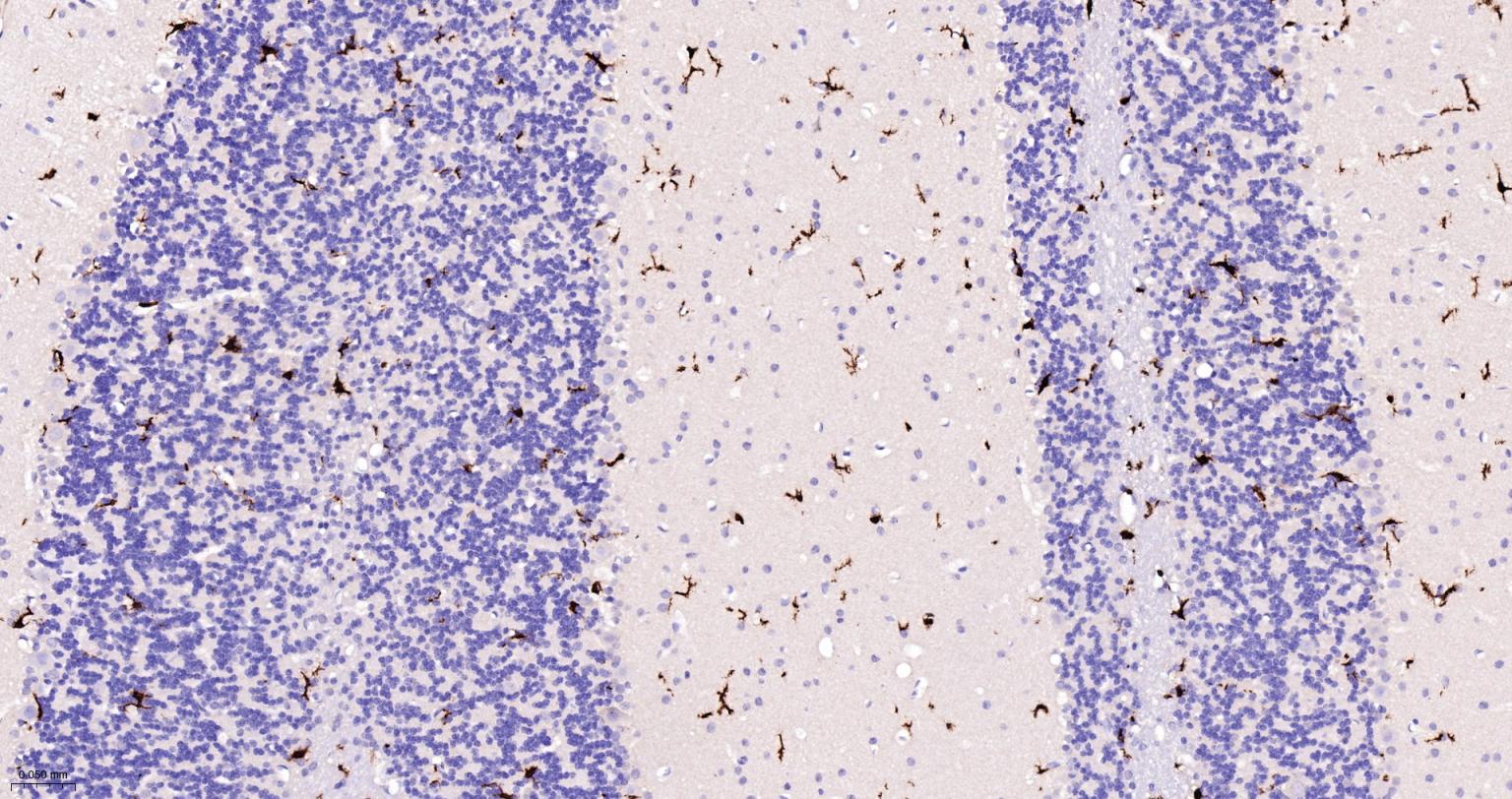
Paraformaldehyde-fixed, paraffin embedded Rat Cerebellum; Antigen retrieval by boiling in sodium citrate buffer (pH6.0) for 15 min; The section was incubated with AIF1 Monoclonal Antibody, Unconjugated (bsm-54132R) at 1:200 overnight at 4°C, followed by conjugation to the bs-0295G-HRP and DAB (C-0010) staining.
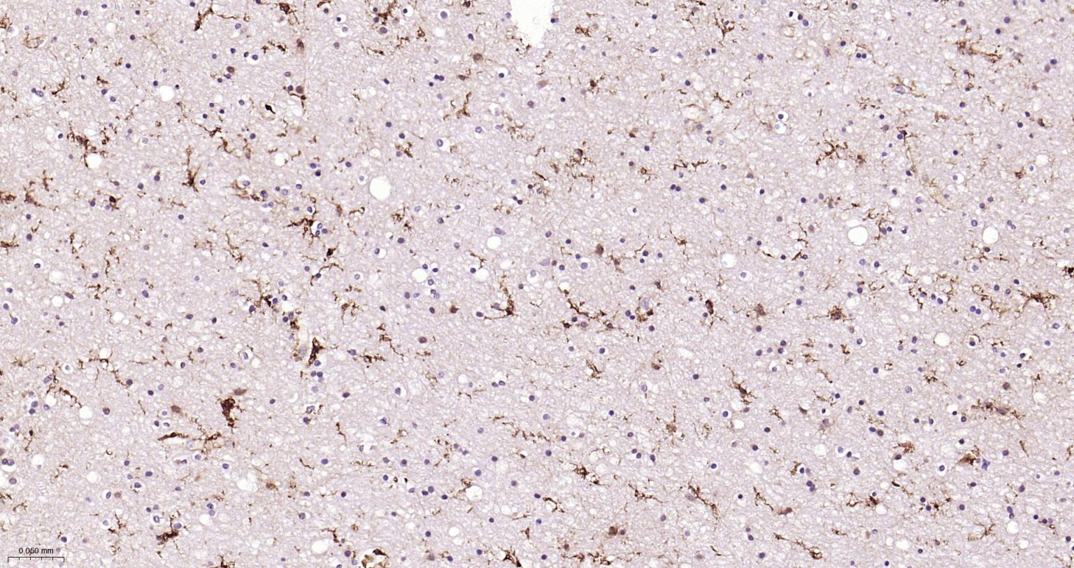
Paraformaldehyde-fixed, paraffin embedded Human Left Parietal Lobe; Antigen retrieval by boiling in sodium citrate buffer (pH6.0) for 15 min; Antibody incubation with AIF1 (9A3) Monoclonal Antibody, Unconjugated(bsm-54132R) at 1:200 overnight at 4°C, followed by conjugation to the SP Kit(Rabbit, SP-0023) and DAB (C-0010) staining.
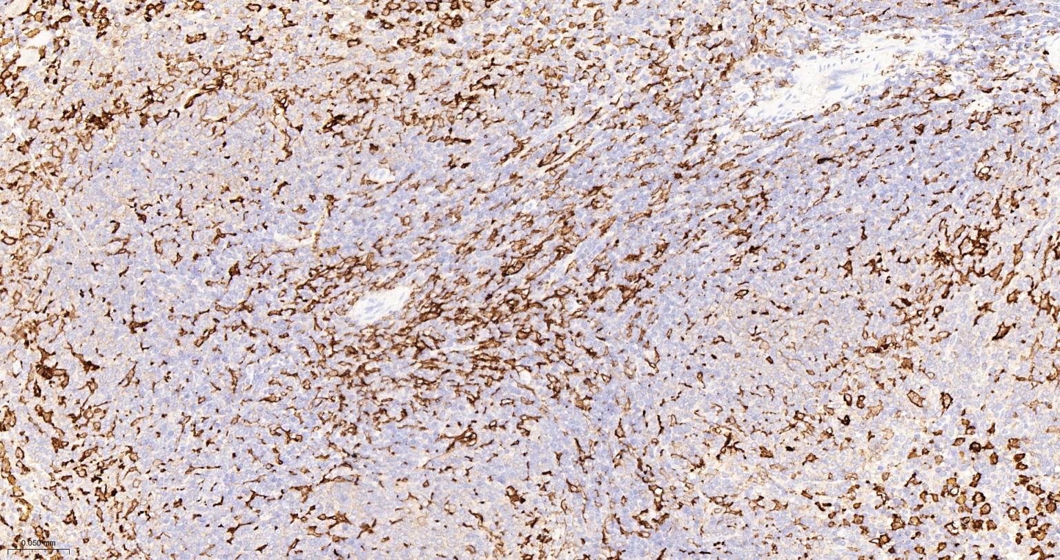
Paraformaldehyde-fixed, paraffin embedded Mouse Spleen; Antigen retrieval by boiling in sodium citrate buffer (pH6.0) for 15 min; Antibody incubation with AIF1 (9A3) Monoclonal Antibody, Unconjugated(bsm-54132R) at 1:500 overnight at 4°C, followed by conjugation to the SP Kit(Rabbit, SP-0023) and DAB (C-0010) staining.
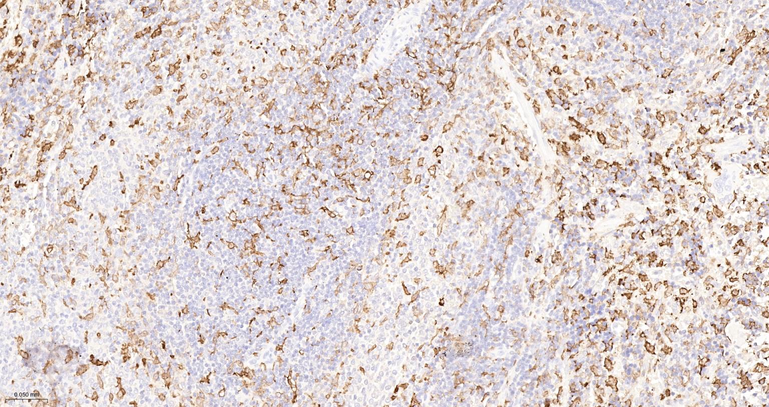
Paraformaldehyde-fixed, paraffin embedded Rat Spleen; Antigen retrieval by boiling in sodium citrate buffer (pH6.0) for 15 min; Antibody incubation with AIF1 (9A3) Monoclonal Antibody, Unconjugated(bsm-54132R) at 1:500 overnight at 4°C, followed by conjugation to the SP Kit(Rabbit, SP-0023) and DAB (C-0010) staining.
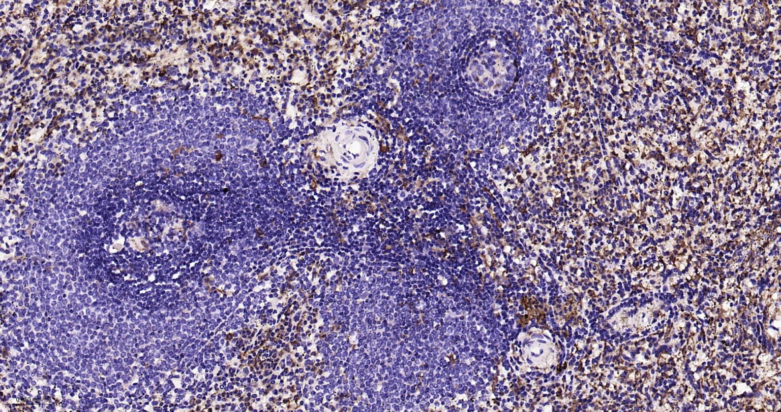
Paraformaldehyde-fixed, paraffin embedded Human Spleen; Antigen retrieval by boiling in sodium citrate buffer (pH6.0) for 15 min; Antibody incubation with AIF1 (9A3) Monoclonal Antibody, Unconjugated(bsm-54132R) at 1:200 overnight at 4°C, followed by conjugation to the SP Kit(Rabbit, SP-0023) and DAB (C-0010) staining.
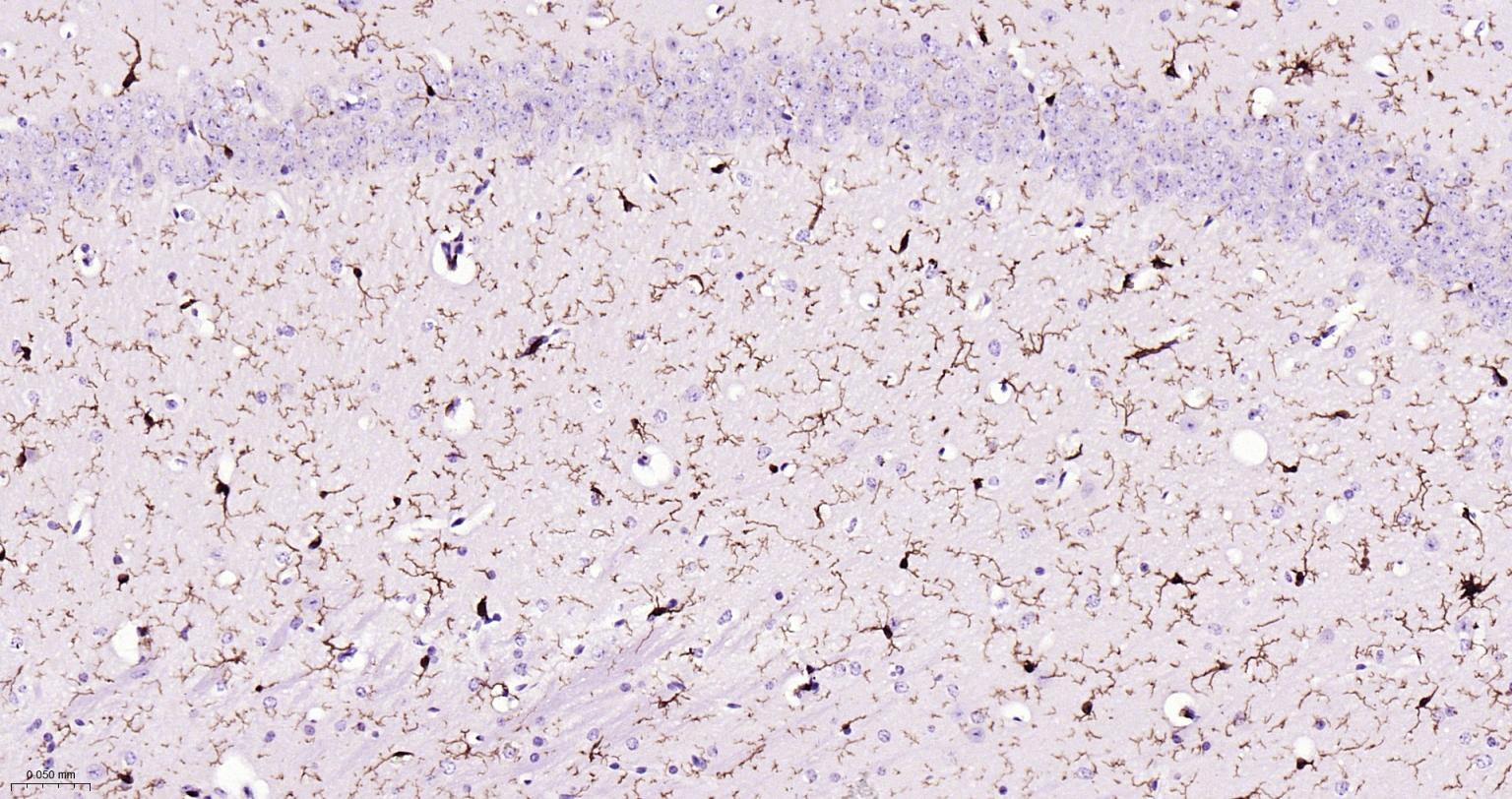
Paraformaldehyde-fixed, paraffin embedded Mouse Cerebrum; Antigen retrieval by boiling in sodium citrate buffer (pH6.0) for 15 min; Antibody incubation with AIF1 (9A3) Monoclonal Antibody, Unconjugated(bsm-54132R) at 1:200 overnight at 4°C, followed by conjugation to the SP Kit(Rabbit, SP-0023) and DAB (C-0010) staining.
Paraformaldehyde-fixed, paraffin embedded Rat Cerebrum; Antigen retrieval by boiling in sodium citrate buffer (pH6.0) for 15 min; Antibody incubation with AIF1 (9A3) Monoclonal Antibody, Unconjugated(bsm-54132R) at 1:200 overnight at 4°C, followed by conjugation to the SP Kit(Rabbit, SP-0023) and DAB (C-0010) staining.
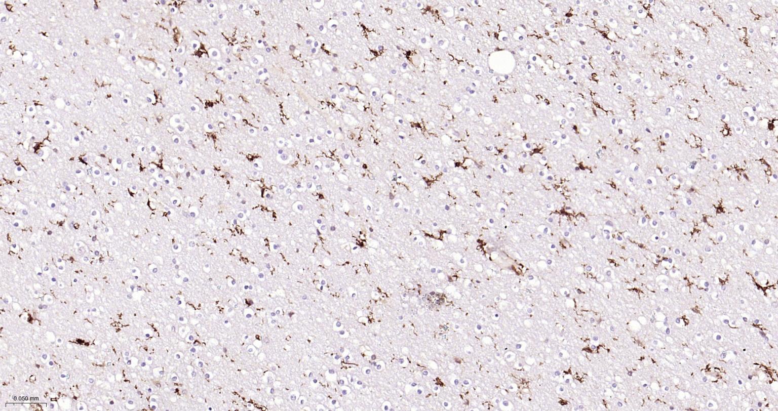
Paraformaldehyde-fixed, paraffin embedded Human Cerebrum; Antigen retrieval by boiling in sodium citrate buffer (pH6.0) for 15 min; Antibody incubation with AIF1 (9A3) Monoclonal Antibody, Unconjugated(bsm-54132R) at 1:200 overnight at 4°C, followed by conjugation to the SP Kit(Rabbit, SP-0023) and DAB (C-0010) staining.
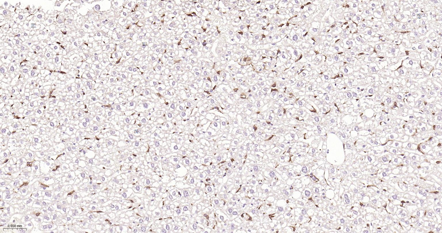
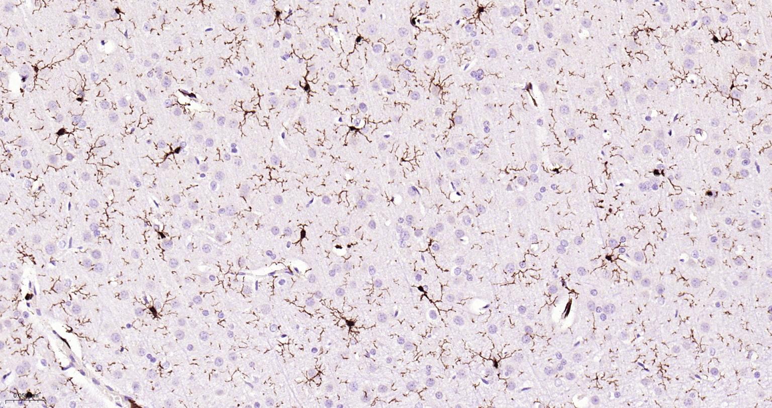
Paraformaldehyde-fixed, paraffin embedded Mouse Liver; Antigen retrieval by boiling in sodium citrate buffer (pH6.0) for 15 min; Antibody incubation with AIF1 (9A3) Monoclonal Antibody, Unconjugated(bsm-54132R) at 1:200 overnight at 4°C, followed by conjugation to the SP Kit(Rabbit, SP-0023) and DAB (C-0010) staining.
Paraformaldehyde-fixed, paraffin embedded Rat Liver; Antigen retrieval by boiling in sodium citrate buffer (pH6.0) for 15 min; Antibody incubation with AIF1 (9A3) Monoclonal Antibody, Unconjugated(bsm-54132R) at 1:200 overnight at 4°C, followed by conjugation to the SP Kit(Rabbit, SP-0023) and DAB (C-0010) staining.
Paraformaldehyde-fixed, paraffin embedded Human Liver; Antigen retrieval by boiling in sodium citrate buffer (pH6.0) for 15 min; Antibody incubation with AIF1 (9A3) Monoclonal Antibody, Unconjugated(bsm-54132R) at 1:200 overnight at 4°C, followed by conjugation to the SP Kit(Rabbit, SP-0023) and DAB (C-0010) staining.
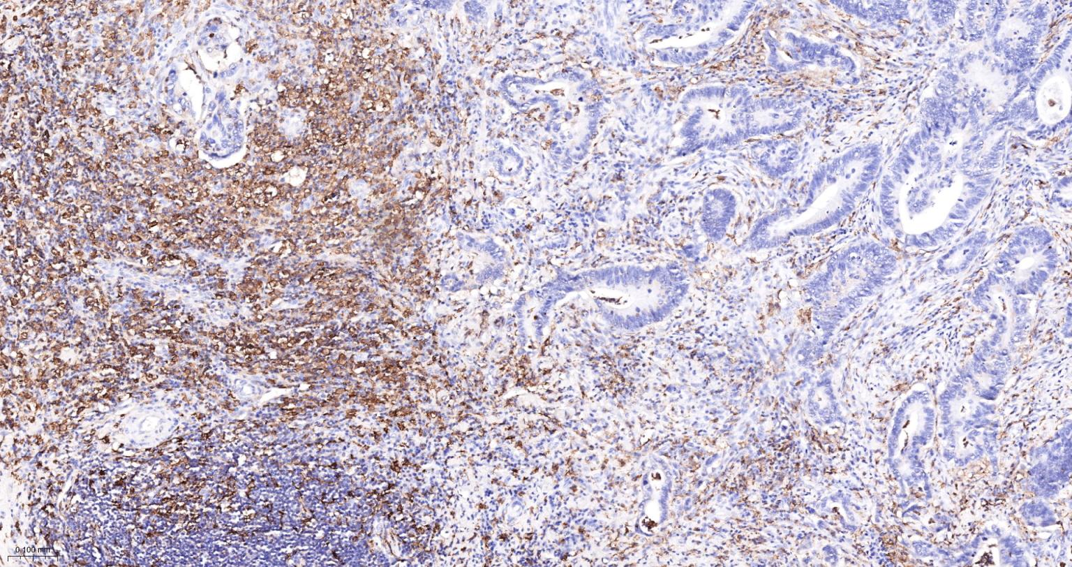
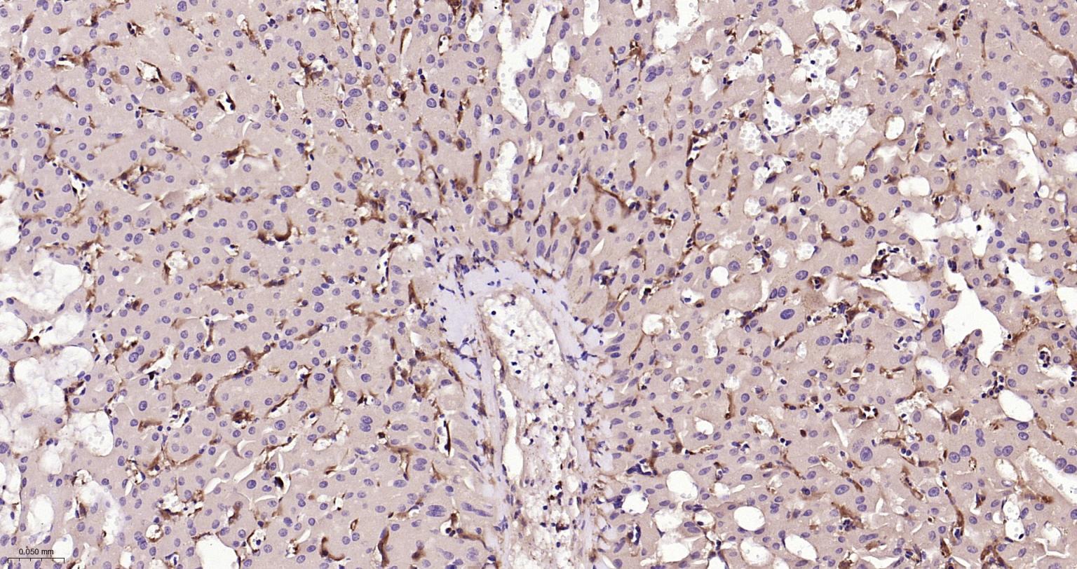
Paraformaldehyde-fixed, paraffin embedded Human Colon Cancer; Antigen retrieval by boiling in sodium citrate buffer (pH6.0) for 15 min; Antibody incubation with AIF1 (9A3) Monoclonal Antibody, Unconjugated(bsm-54132R) at 1:500 overnight at 4°C, followed by conjugation to the SP Kit(Rabbit, SP-0023) and DAB (C-0010) staining.
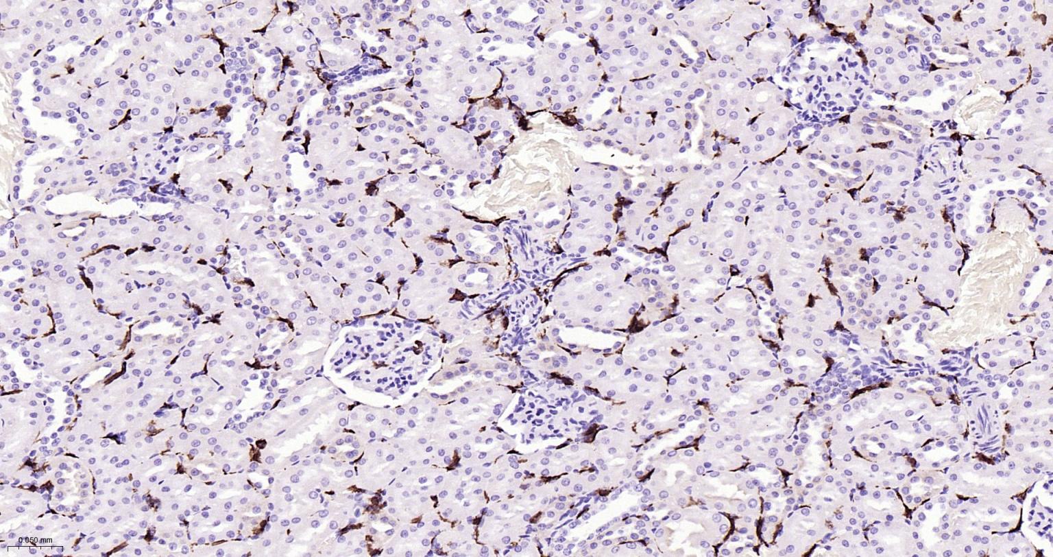
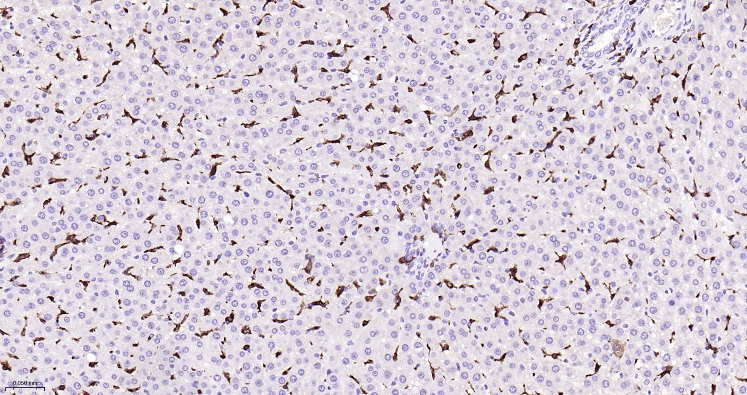
Paraformaldehyde-fixed, paraffin embedded Rat Kidney; Antigen retrieval by boiling in sodium citrate buffer (pH6.0) for 15 min; Antibody incubation with AIF1 (9A3) Monoclonal Antibody, Unconjugated(bsm-54132R) at 1:200 overnight at 4°C, followed by conjugation to the SP Kit(Rabbit, SP-0023) and DAB (C-0010) staining.
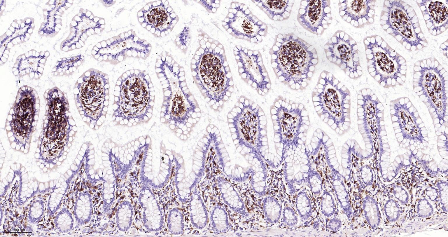
Paraformaldehyde-fixed, paraffin embedded Human Small Intestine; Antigen retrieval by boiling in sodium citrate buffer (pH6.0) for 15 min; Antibody incubation with AIF1 (9A3) Monoclonal Antibody, Unconjugated(bsm-54132R) at 1:200 overnight at 4°C, followed by conjugation to the SP Kit(Rabbit, SP-0023) and DAB (C-0010) staining.
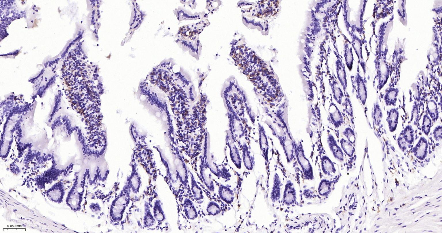
Paraformaldehyde-fixed, paraffin embedded Mouse Small Intestine; Antigen retrieval by boiling in sodium citrate buffer (pH6.0) for 15 min; Antibody incubation with AIF1 (9A3) Monoclonal Antibody, Unconjugated(bsm-54132R) at 1:200 overnight at 4°C, followed by conjugation to the SP Kit(Rabbit, SP-0023) and DAB (C-0010) staining.
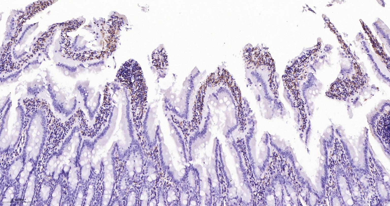
Paraformaldehyde-fixed, paraffin embedded Rat Small Intestine; Antigen retrieval by boiling in sodium citrate buffer (pH6.0) for 15 min; Antibody incubation with AIF1 (9A3) Monoclonal Antibody, Unconjugated(bsm-54132R) at 1:200 overnight at 4°C, followed by conjugation to the SP Kit(Rabbit, SP-0023) and DAB (C-0010) staining.
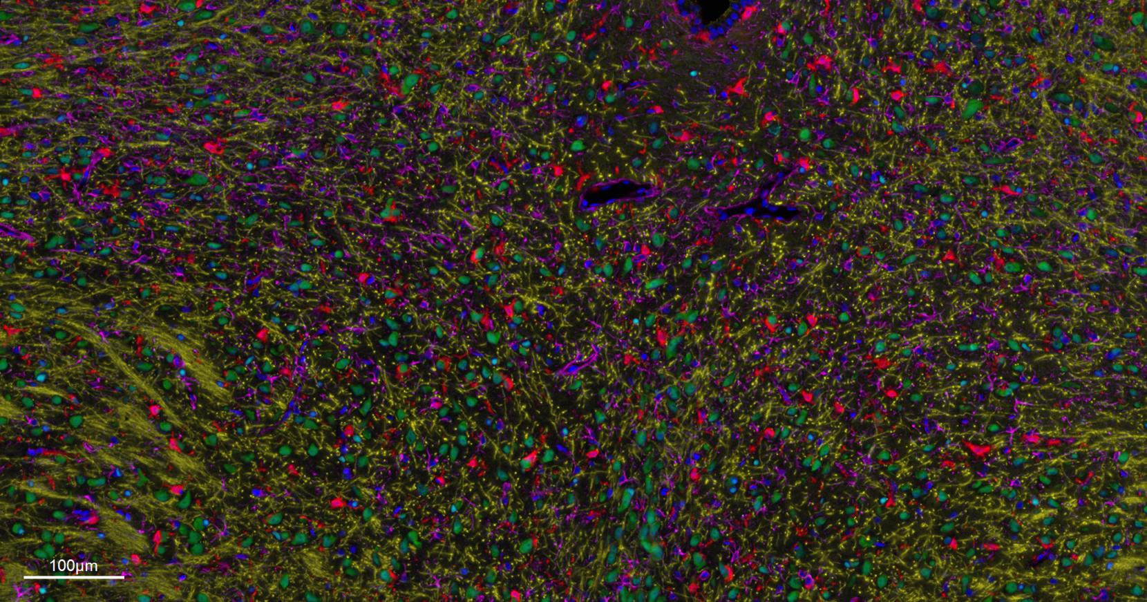
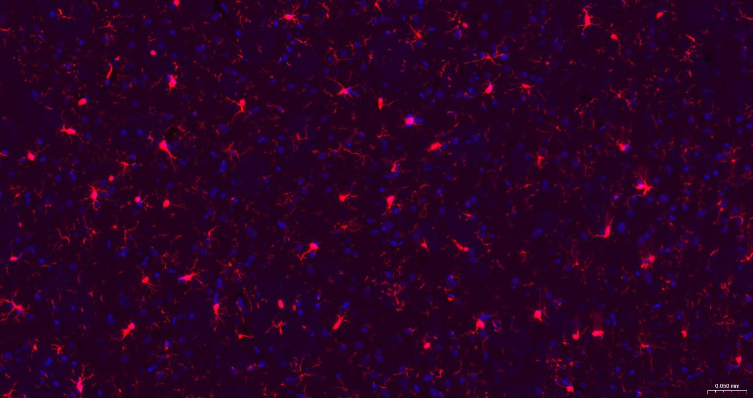
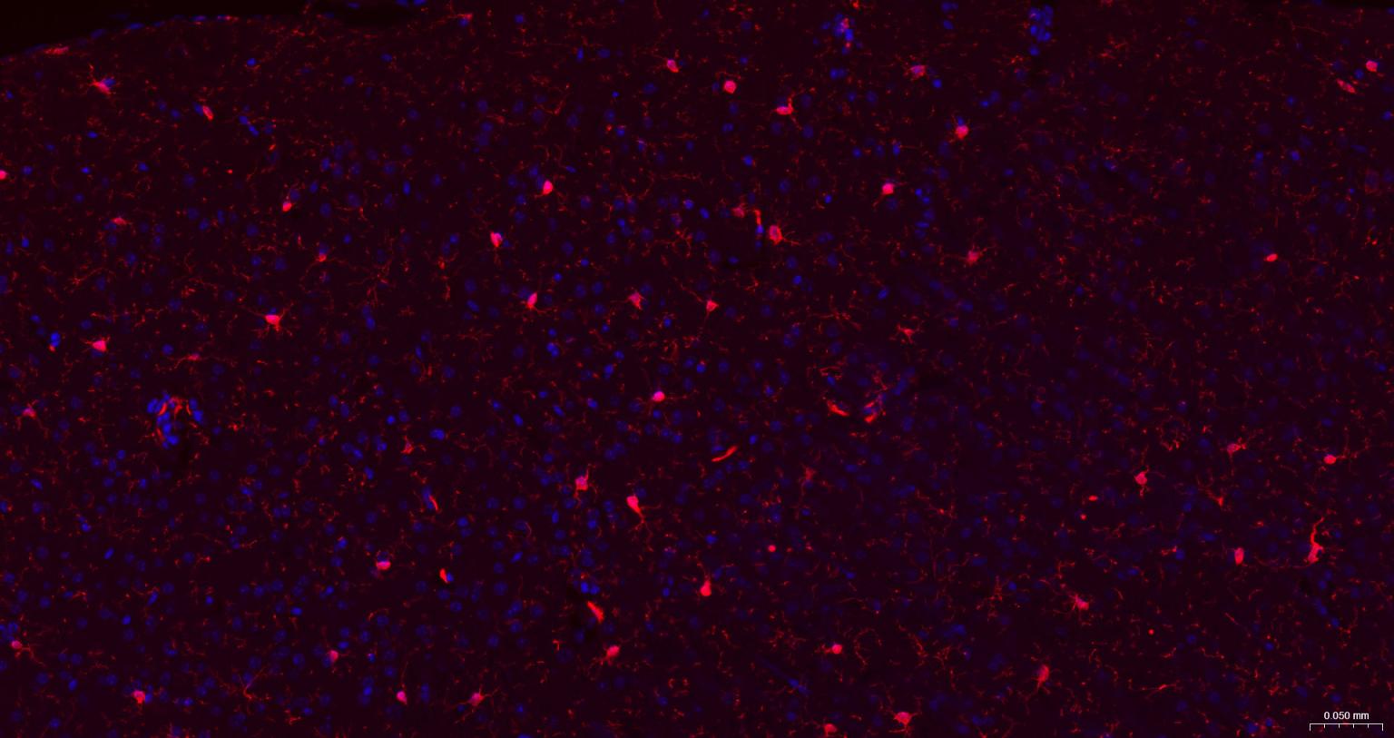
Paraformaldehyde-fixed, paraffin embedded Mouse Cerebrum.
Merged staining of anti-GFAP (bsm-42001R; 1:200; pink) anti-AIF1/Iba1 (bsm-54132R; 1:200; red) anti-NeuN (bsm-52268R; 1:200; green) anti-MBP (bsm-33932M; 1:200; yellow) and anti-Olig2 (bsm-61115R; 1:200; light blue) .
DAPI (dark blue) was used as a nuclear counter stain.
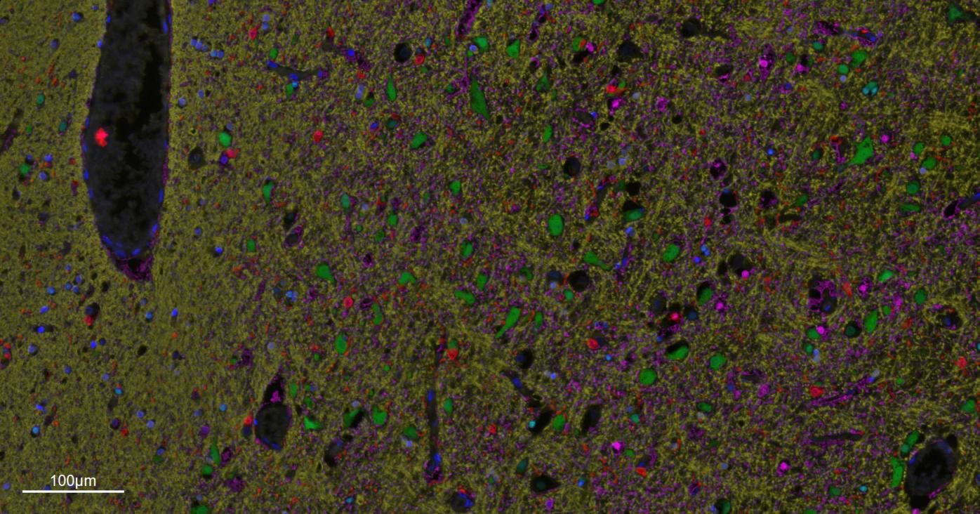
Paraformaldehyde-fixed, paraffin embedded Human Cerebrum.
Merged staining of anti-GFAP (bsm-42001R; 1:200; pink) anti-AIF1/Iba1 (bsm-54132R; 1:200; red) anti-NeuN (bsm-52268R; 1:200; green) anti-MBP (bsm-33932M; 1:200; yellow) and anti-Olig2 (bsm-61115R; 1:200; light blue) .
DAPI (dark blue) was used as a nuclear counter stain.
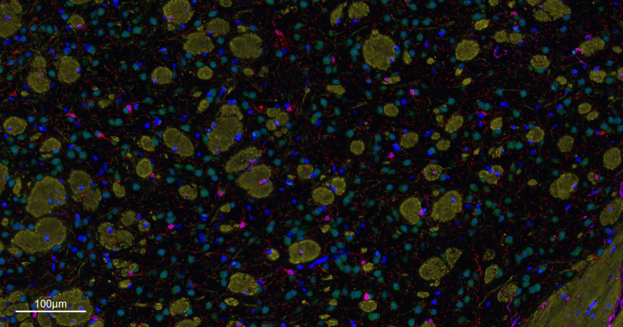
Paraformaldehyde-fixed, paraffin embedded Rat Cerebrum.
Merged staining of anti-GFAP (bsm-42001R; 1:200; pink) anti-AIF1/Iba1 (bsm-54132R; 1:200; red) anti-NeuN (bsm-52268R; 1:200; green) anti-MBP (bsm-33932M; 1:200; yellow) and anti-Olig2 (bsm-61115R; 1:200; light blue) .
DAPI (dark blue) was used as a nuclear counter stain.
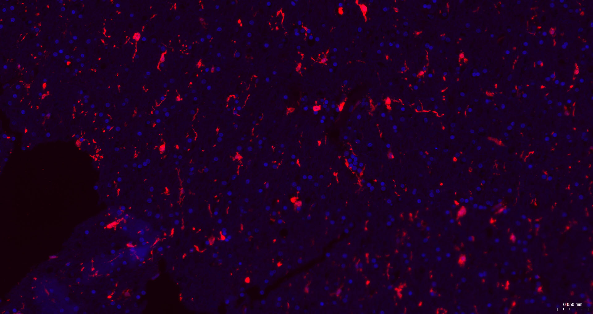
Paraformaldehyde-fixed, paraffin embedded Human left parietal lobe; Antigen retrieval by boiling in sodium citrate buffer (pH6.0) for 15 min; The section was incubated with AIF1 Monoclonal Antibody, Unconjugated (bsm-54132R) at 1:200 overnight at 4°C. Followed by conjugated Goat Anti-Rabbit IgG antibody (Red, bs-0295G-BF594), DAPI (blue, C02-04002) was used to stain the cell nuclei.
Paraformaldehyde-fixed, paraffin embedded Rat Cerebrum; Antigen retrieval by boiling in sodium citrate buffer (pH6.0) for 15 min; The section was incubated with AIF1 Monoclonal Antibody, Unconjugated (bsm-54132R) at 1:200 overnight at 4°C. Followed by conjugated Goat Anti-Rabbit IgG antibody (Red, bs-0295G-BF594), DAPI (blue, C02-04002) was used to stain the cell nuclei.
Paraformaldehyde-fixed, paraffin embedded Mouse Cerebrum; Antigen retrieval by boiling in sodium citrate buffer (pH6.0) for 15 min; The section was incubated with AIF1 Monoclonal Antibody, Unconjugated (bsm-54132R) at 1:200 overnight at 4°C. Followed by conjugated Goat Anti-Rabbit IgG antibody (Red, bs-0295G-BF594), DAPI (blue, C02-04002) was used to stain the cell nuclei.
|
| 1、抗体溶解方法 | |
| 2、抗体修复方式 | |
| 3、常用试剂的配制 | |
| 4、免疫组化操作步骤 | |
| 5、免疫组化问题解答 | |
| 6、Western Blotting 操作步骤 | |
| 7、Western Blotting 问题解答 | |
| 8、关于肽链的设计 | |
| 9、多肽的溶解与保存 | |
| 10、酶标抗体效价测定程序 | |


