
| 产品编号 | bsm-54176R |
| 英文名称 | Histone H1.2 Recombinant Rabbit mAb |
| 中文名称 | 组蛋白H1.2重组兔单抗 |
| 别 名 | H1.2; H1C; H1F2; H1s-1; HIST1H1C; H12_HUMAN; H1-2; Histone H1c; Histone H1d; Histone H1s-1; H1.2 linker histone, cluster member; H1 histone family, member 2; histone 1, H1c; histone cluster 1, H1c; histone cluster 1 H1 family member c |
| 抗体来源 | Rabbit |
| 克隆类型 | Recombinant |
| 克 隆 号 | 9C9 |
| 交叉反应 | Human,Mouse,Rat |
| 产品应用 | WB=1:500-2000,Flow-Cyt=1:50-100,ICC/IF=1:50-200
not yet tested in other applications. optimal dilutions/concentrations should be determined by the end user. |
| 理论分子量 | 21kDa |
| 检测分子量 | 32 |
| 细胞定位 | 细胞核 |
| 性 状 | Liquid |
| 浓 度 | 1mg/ml |
| 免 疫 原 | KLH conjugated synthetic peptide derived from human Histone H1.2 |
| 亚 型 | IgG |
| 纯化方法 | affinity purified by Protein A |
| 缓 冲 液 | 0.01M TBS (pH7.4) with 1% BSA, 0.02% Proclin300 and 50% Glycerol. |
| 保存条件 | Shipped at 4℃. Store at -20℃ for one year. Avoid repeated freeze/thaw cycles. |
| 注意事项 | This product as supplied is intended for research use only, not for use in human, therapeutic or diagnostic applications. |
| PubMed | PubMed |
| 产品介绍 |
Histones are basic nuclear proteins responsible for nucleosome structure of the chromosomal fiber in eukaryotes. Two molecules of each of the four core histones (H2A, H2B, H3, and H4) form an octamer, around which approximately 146 bp of DNA is wrapped in repeating units, called nucleosomes. The linker histone, H1, interacts with linker DNA between nucleosomes and functions in the compaction of chromatin into higher order structures. This gene is intronless and encodes a replication-dependent histone that is a member of the histone H1 family. Transcripts from this gene lack polyA tails but instead contain a palindromic termination element. This gene is found in the large histone gene cluster on chromosome 6. [provided by RefSeq, Aug 2015] Function: Histone H1 protein binds to linker DNA between nucleosomes forming the macromolecular structure known as the chromatin fiber. Histones H1 are necessary for the condensation of nucleosome chains into higher-order structured fibers. Acts also as a regulator of individual gene transcription through chromatin remodeling, nucleosome spacing and DNA methylation (By similarity). Subcellular Location: Nucleus. Tissue Specificity: Expressed in calcaneal tendon and 216 other tissues. Post-translational modifications: H1 histones are progressively phosphorylated during the cell cycle, becoming maximally phosphorylated during late G2 phase and M phase, and being dephosphorylated sharply thereafter. Crotonylation (Kcr) is specifically present in male germ cells and marks testis-specific genes in post-meiotic cells, including X-linked genes that escape sex chromosome inactivation in haploid cells. Crotonylation marks active promoters and enhancers and confers resistance to transcriptional repressors. It is also associated with post-meiotically activated genes on autosomes. Citrullination at Arg-54 (H1R54ci) by PADI4 takes place within the DNA-binding site of H1 and results in its displacement from chromatin and global chromatin decondensation, thereby promoting pluripotency and stem cell maintenance. ADP-ribosylated on Ser-188 in response to DNA damage. SWISS: P16403 Gene ID: 3006 Database links: Entrez Gene: 3006 Human Omim: 142710 Human SwissProt: P16403 Human Unigene: 7644 Human |
| 产品图片 |
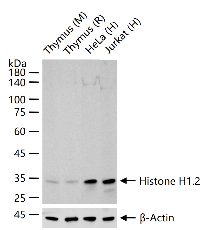
25 ug total protein per lane of various lysates (see on figure) probed with Histone H1.2 monoclonal antibody, unconjugated (bsm-54176R) at 1:1000 dilution and 4°C overnight incubation. Followed by conjugated secondary antibody incubation at r.t. for 60 min.
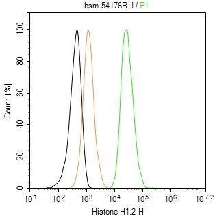
The HeLa (H) cells were fixed with 4% PFA (10 min at r.t.) and then permeabilized with 90% ice-cold methanol for 20 min at -20℃,the cells then were incubated in 5%BSA to block non-specific protein-protein interactions (30 min at r.t.), followed by secondary antibody incubation for 40 min at room temperature. Primary Antibody (green):Rabbit Anti-Histone H1.2 antibody (bsm-54176R,1:100); Isotype Control (orange): Rabbit IgG (bs-0295P). Blank control (black): PBS. Acquisition of 20,000 events was performed.
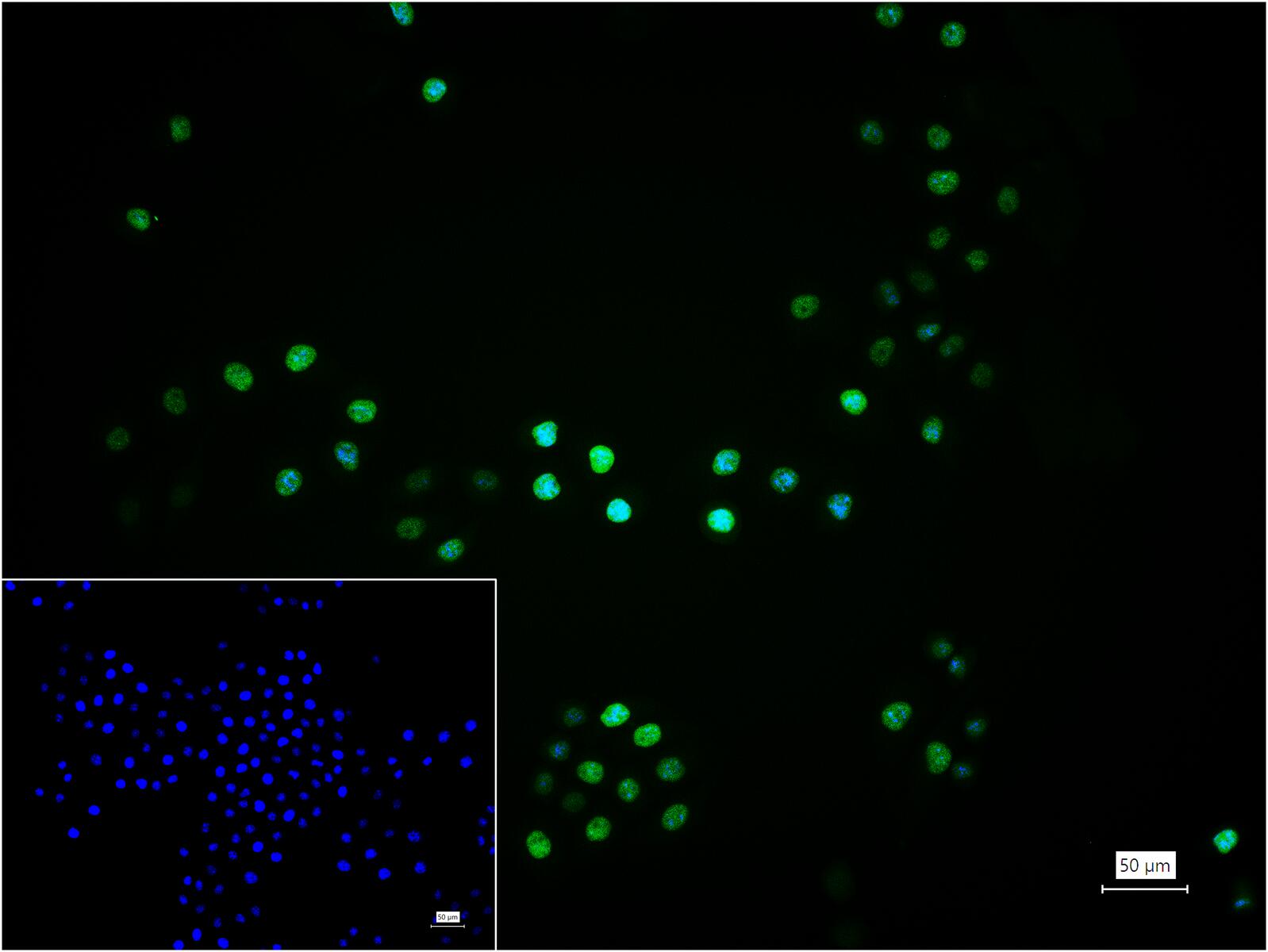
4% Paraformaldehyde-fixed HeLa (H) cell; Triton X-100 at r.t. for 20 min; Antibody incubation with (Histone H1.2) monoclonal Antibody, unconjugated (bsm-54176R) 1:100, 90 min at 37°C; followed by BF488 conjugated Goat Anti-Rabbit IgG antibody (green, bs-60295G-BF488) at 37°C for 90 min, DAPI (blue, C02-04002) was used to stain the cell nuclei. PBS instead of the primary antibody was used as the blank control.
|
| 1、抗体溶解方法 | |
| 2、抗体修复方式 | |
| 3、常用试剂的配制 | |
| 4、免疫组化操作步骤 | |
| 5、免疫组化问题解答 | |
| 6、Western Blotting 操作步骤 | |
| 7、Western Blotting 问题解答 | |
| 8、关于肽链的设计 | |
| 9、多肽的溶解与保存 | |
| 10、酶标抗体效价测定程序 | |


