
| 产品编号 | bsm-54085R |
| 英文名称 | TGFBI Recombinant Rabbit mAb |
| 中文名称 | 角膜上皮蛋白TGFBI重组兔单抗 |
| 别 名 | BIGH3; CDB1; CDG2; CDGG1; CSD; CSD1; CSD2; CSD3; EBMD; LCD1; 68kDa; Beta-ig; big-h3; BGH3_HUMAN; TGFBI; Beta ig-h3; Kerato-epithelin; RGD-containing collagen-associated protein (RGD-CAP); BGH3_MOUSE; |
| 研究领域 | 细胞生物 发育生物学 神经生物学 信号转导 干细胞 生长因子和激素 |
| 抗体来源 | Rabbit |
| 克隆类型 | Recombinant |
| 克 隆 号 | 9C2 |
| 交叉反应 | Human,Mouse,Rat |
| 产品应用 | WB=1:500-2000,IHC-P=1:50-200,IHC-F=1:50-200,IF=1:50-200,Flow-Cyt=1:50-100
not yet tested in other applications. optimal dilutions/concentrations should be determined by the end user. |
| 理论分子量 | 72 kDa |
| 检测分子量 | 65 |
| 细胞定位 | 分泌型蛋白 |
| 性 状 | Liquid |
| 浓 度 | 1mg/ml |
| 免 疫 原 | A synthesized peptide derived from human TGFBI: 100-150 |
| 亚 型 | IgG |
| 纯化方法 | affinity purified by Protein A |
| 缓 冲 液 | 0.01M TBS (pH7.4) with 1% BSA, 0.02% Proclin300 and 50% Glycerol. |
| 保存条件 | Shipped at 4℃. Store at -20℃ for one year. Avoid repeated freeze/thaw cycles. |
| 注意事项 | This product as supplied is intended for research use only, not for use in human, therapeutic or diagnostic applications. |
| PubMed | PubMed |
| 产品介绍 |
This gene encodes an RGD-containing protein that binds to type I, II and IV collagens. The RGD motif is found in many extracellular matrix proteins modulating cell adhesion and serves as a ligand recognition sequence for several integrins. This protein plays a role in cell-collagen interactions and may be involved in endochondrial bone formation in cartilage. The protein is induced by transforming growth factor-beta and acts to inhibit cell adhesion. Mutations in this gene are associated with multiple types of corneal dystrophy. [provided by RefSeq, Jul 2008] Function: Binds to type I, II, and IV collagens. This adhesion protein may play an important role in cell-collagen interactions. In cartilage, may be involved in endochondral bone formation. Subcellular Location: Secreted > extracellular space > extracellular matrix. May be associated both with microfibrils and with the cell surface. Tissue Specificity : Highly expressed in the corneal epithelium. Post-translational modifications: Gamma-carboxyglutamate residues are formed by vitamin K dependent carboxylation. These residues are essential for the binding of calcium. DISEASE: Defects in TGFBI are the cause of epithelial basement membrane corneal dystrophy (EBMD) [MIM:121820]; also known as Cogan corneal dystrophy or map-dot-fingerprint type corneal dystrophy. EBMD is a bilateral anterior corneal dystrophy characterized by grayish epithelial fingerprint lines, geographic map-like lines, and dots (or microcysts) on slit-lamp examination. Pathologic studies show abnormal, redundant basement membrane and intraepithelial lacunae filled with cellular debris. Although this disorder usually is not considered to be inherited, families with autosomal dominant inheritance have been identified. Defects in TGFBI are the cause of corneal dystrophy Groenouw type 1 (CDGG1) [MIM:121900]; also known as corneal dystrophy granular type. Inheritance is autosomal dominant. Corneal dystrophies show progressive opacification of the cornea leading to severe visual handicap. Defects in TGFBI are the cause of corneal dystrophy lattice type 1 (CDL1) [MIM:122200]. Inheritance is autosomal dominant. Defects in TGFBI are a cause of corneal dystrophy Thiel-Behnke type (CDTB) [MIM:602082]; also known as corneal dystrophy of Bowman layer type 2 (CDB2). Defects in TGFBI are the cause of Reis-Buecklers corneal dystrophy (CDRB) [MIM:608470]; also known as corneal dystrophy of Bowman layer type 1 (CDB1). Defects in TGFBI are the cause of lattice corneal dystrophy type 3A (CDL3A) [MIM:608471]. CDL3A clinically resembles to lattice corneal dystrophy type 3, but differs in that its age of onset is 70 to 90 years. It has an autosomal dominant inheritance pattern. Defects in TGFBI are the cause of Avellino corneal dystrophy (ACD) [MIM:607541]. ACD could be considered a variant of granular dystrophy with a significant amyloidogenic tendency. Inheritance is autosomal dominant. Similarity: Contains 1 EMI domain. Contains 4 FAS1 domains. SWISS: Q15582 Gene ID: 7045 |
| 产品图片 |
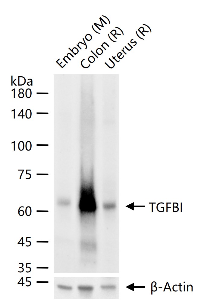
25 ug total protein per lane of various lysates (see on figure) probed with TGFBI monoclonal antibody, unconjugated (bsm-54085R) at 1:1000 dilution and 4°C overnight incubation. Followed by conjugated secondary antibody incubation at r.t. for 60 min.
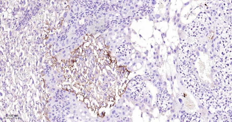
Paraformaldehyde-fixed, paraffin embedded Human Colon Cancer; Antigen retrieval by boiling in sodium citrate buffer (pH6.0) for 15 min; The section was incubated with TGFBI Monoclonal Antibody, Unconjugated (bsm-54085R) at 1:200 overnight at 4°C, followed by conjugation to the bs-0295G-HRP and DAB (C-0010) staining.
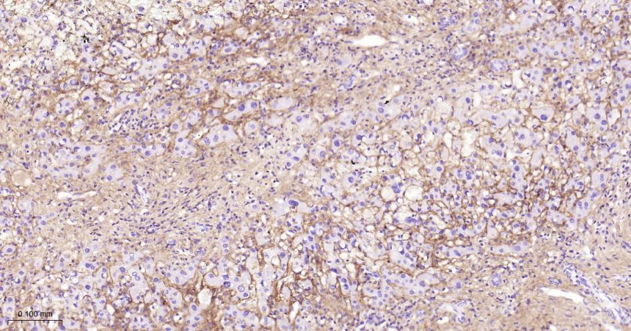
Paraformaldehyde-fixed, paraffin embedded Human Liver Cancer; Antigen retrieval by boiling in sodium citrate buffer (pH6.0) for 15 min; The section was incubated with TGFBI Monoclonal Antibody, Unconjugated (bsm-54085R) at 1:200 overnight at 4°C, followed by conjugation to the bs-0295G-HRP and DAB (C-0010) staining.
Paraformaldehyde-fixed, paraffin embedded Human Lung Cancer; Antigen retrieval by boiling in sodium citrate buffer (pH6.0) for 15 min; The section was incubated with TGFBI Monoclonal Antibody, Unconjugated (bsm-54085R) at 1:200 overnight at 4°C, followed by conjugation to the bs-0295G-HRP and DAB (C-0010) staining.
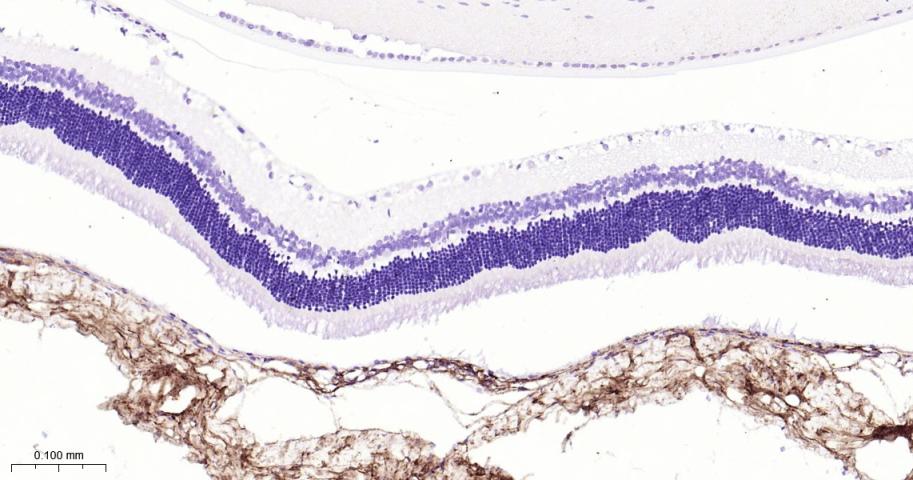
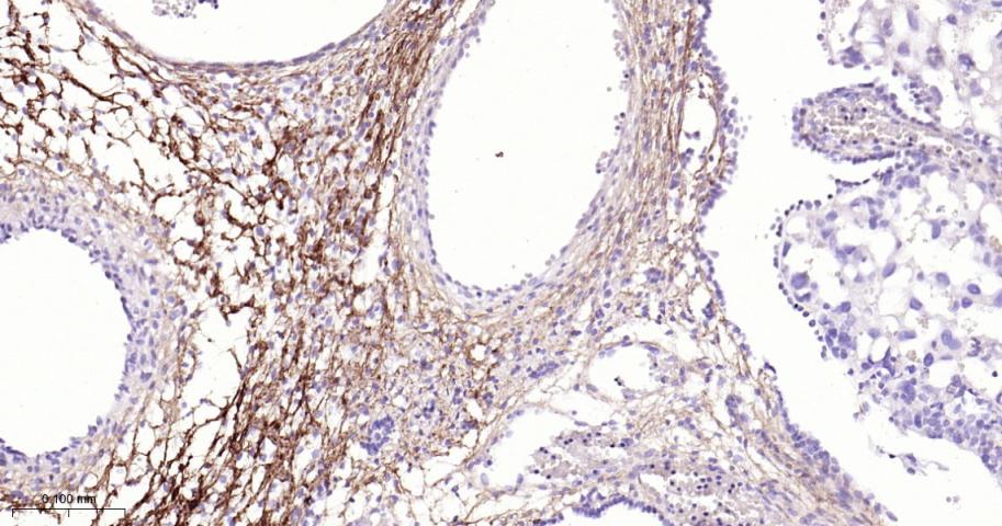
Paraformaldehyde-fixed, paraffin embedded Rat Eye; Antigen retrieval by boiling in sodium citrate buffer (pH6.0) for 15 min; The section was incubated with TGFBI Monoclonal Antibody, Unconjugated (bsm-54085R) at 1:200 overnight at 4°C, followed by conjugation to the bs-0295G-HRP and DAB (C-0010) staining.
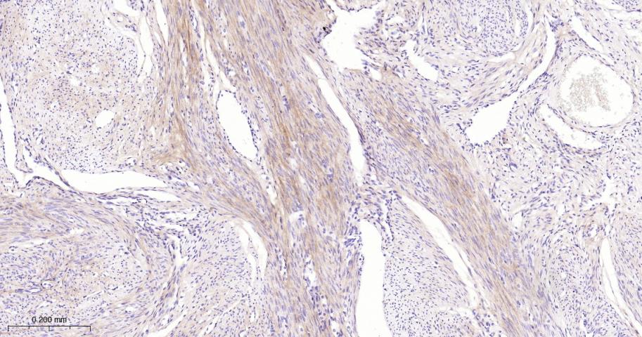
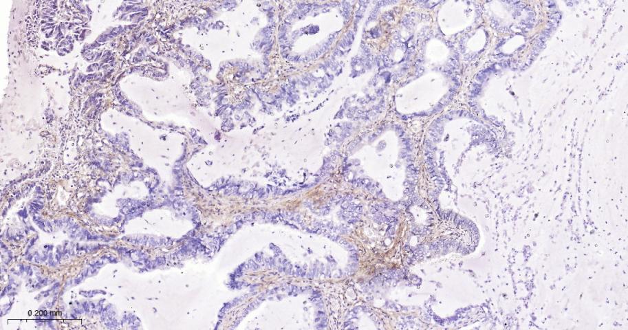
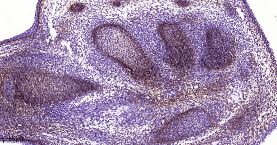
Paraformaldehyde-fixed, paraffin embedded Human Uterus; Antigen retrieval by boiling in sodium citrate buffer (pH6.0) for 15 min; The section was incubated with TGFBI Monoclonal Antibody, Unconjugated (bsm-54085R) at 1:200 overnight at 4°C, followed by conjugation to the bs-0295G-HRP and DAB (C-0010) staining.
Paraformaldehyde-fixed, paraffin embedded Mouse Uterus; Antigen retrieval by boiling in sodium citrate buffer (pH6.0) for 15 min; The section was incubated with TGFBI Monoclonal Antibody, Unconjugated (bsm-54085R) at 1:200 overnight at 4°C, followed by conjugation to the bs-0295G-HRP and DAB (C-0010) staining.
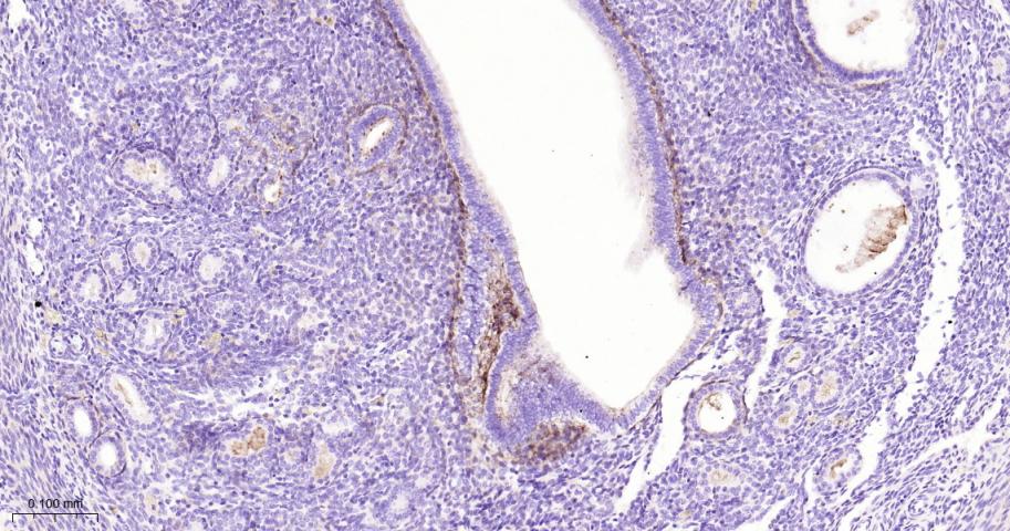
Paraformaldehyde-fixed, paraffin embedded Human Placenta; Antigen retrieval by boiling in sodium citrate buffer (pH6.0) for 15 min; The section was incubated with TGFBI Monoclonal Antibody, Unconjugated (bsm-54085R) at 1:200 overnight at 4°C, followed by conjugation to the bs-0295G-HRP and DAB (C-0010) staining.
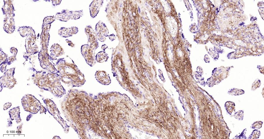
Paraformaldehyde-fixed, paraffin embedded Rat Placenta; Antigen retrieval by boiling in sodium citrate buffer (pH6.0) for 15 min; The section was incubated with TGFBI Monoclonal Antibody, Unconjugated (bsm-54085R) at 1:200 overnight at 4°C, followed by conjugation to the bs-0295G-HRP and DAB (C-0010) staining.
Paraformaldehyde-fixed, paraffin embedded Mouse Placenta; Antigen retrieval by boiling in sodium citrate buffer (pH6.0) for 15 min; The section was incubated with TGFBI Monoclonal Antibody, Unconjugated (bsm-54085R) at 1:200 overnight at 4°C, followed by conjugation to the bs-0295G-HRP and DAB (C-0010) staining.

Paraformaldehyde-fixed, paraffin embedded Rat Embryo; Antigen retrieval by boiling in sodium citrate buffer (pH6.0) for 15 min; The section was incubated with TGFBI Monoclonal Antibody, Unconjugated (bsm-54085R) at 1:200 overnight at 4°C, followed by conjugation to the bs-0295G-HRP and DAB (C-0010) staining.
Paraformaldehyde-fixed, paraffin embedded Mouse Embryo; Antigen retrieval by boiling in sodium citrate buffer (pH6.0) for 15 min; The section was incubated with TGFBI Monoclonal Antibody, Unconjugated (bsm-54085R) at 1:200 overnight at 4°C, followed by conjugation to the bs-0295G-HRP and DAB (C-0010) staining.
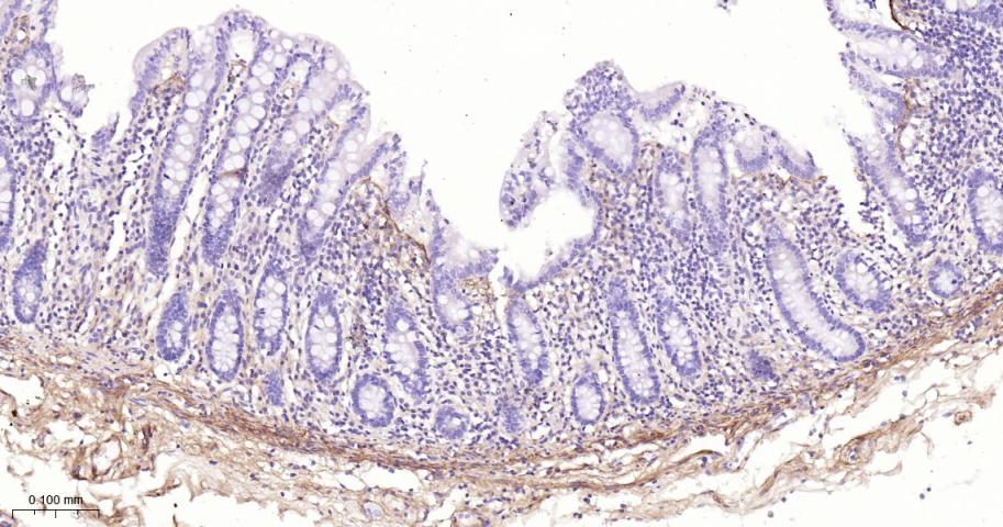
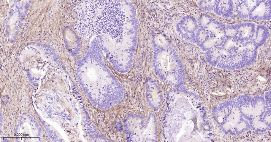
Paraformaldehyde-fixed, paraffin embedded Human Colon; Antigen retrieval by boiling in sodium citrate buffer (pH6.0) for 15 min; The section was incubated with TGFBI Monoclonal Antibody, Unconjugated (bsm-54085R) at 1:200 overnight at 4°C, followed by conjugation to the bs-0295G-HRP and DAB (C-0010) staining.
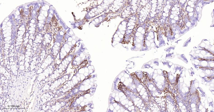
Paraformaldehyde-fixed, paraffin embedded Rat Colon; Antigen retrieval by boiling in sodium citrate buffer (pH6.0) for 15 min; The section was incubated with TGFBI Monoclonal Antibody, Unconjugated (bsm-54085R) at 1:200 overnight at 4°C, followed by conjugation to the bs-0295G-HRP and DAB (C-0010) staining.
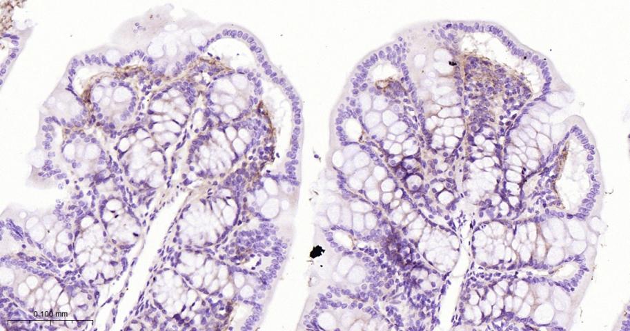
Paraformaldehyde-fixed, paraffin embedded Mouse Colon; Antigen retrieval by boiling in sodium citrate buffer (pH6.0) for 15 min; The section was incubated with TGFBI Monoclonal Antibody, Unconjugated (bsm-54085R) at 1:200 overnight at 4°C, followed by conjugation to the bs-0295G-HRP and DAB (C-0010) staining.
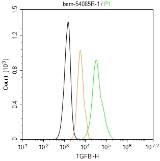
The Hela (H) cells were fixed with 4% PFA (10 min at r.t.) and then permeabilized with 90% ice-cold methanol for 20 min at -20℃,the cells then were incubated in 5%BSA to block non-specific protein-protein interactions (30 min at r.t.), followed by secondary antibody incubation for 40 min at room temperature. Primary Antibody (green):Rabbit Anti-TGFBI antibody (bsm-54085R,1:100); Isotype Control (orange): Rabbit IgG (bs-0295P). Blank control (black): PBS. Acquisition of 20,000 events was performed.
|
| 1、抗体溶解方法 | |
| 2、抗体修复方式 | |
| 3、常用试剂的配制 | |
| 4、免疫组化操作步骤 | |
| 5、免疫组化问题解答 | |
| 6、Western Blotting 操作步骤 | |
| 7、Western Blotting 问题解答 | |
| 8、关于肽链的设计 | |
| 9、多肽的溶解与保存 | |
| 10、酶标抗体效价测定程序 | |


