
| 产品编号 | bsm-52324R |
| 英文名称 | ERG Recombinant Rabbit mAb |
| 中文名称 | 癌基因ERG重组兔单抗 |
| 别 名 | LMPHM14; erg-3; p55; D030036I24Rik; ERG_HUMAN; ERG; Transforming protein ERG; ERG_MOUSE; ETS transcription factor ERG; v-ets avian erythroblastosis virus E26 oncogene related; v-ets avian erythroblastosis virus E26 oncogene homolog; transcriptional regulator ERG (transforming protein ERG); v-ets erythroblastosis virus E26 oncogene like; TMPRSS2-ERG prostate cancer specific; ETS-related gene |
| 研究领域 | 肿瘤 细胞生物 信号转导 转录调节因子 表观遗传学 |
| 抗体来源 | Rabbit |
| 克隆类型 | Recombinant |
| 克 隆 号 | 2F3 |
| 交叉反应 | Human,Mouse,Rat |
| 产品应用 | WB=1:500-2000,IHC-P=1:100-500,IHC-F=1:100-500,IF=1:100-500,Flow-Cyt=1:50-100,ICC/IF=1:50-200
not yet tested in other applications. optimal dilutions/concentrations should be determined by the end user. |
| 理论分子量 | 55kDa |
| 检测分子量 | 60 |
| 细胞定位 | 细胞核 细胞浆 |
| 性 状 | Liquid |
| 浓 度 | 1mg/ml |
| 免 疫 原 | A synthesized peptide derived from human ERG: 400-479 |
| 亚 型 | IgG |
| 纯化方法 | affinity purified by Protein A |
| 缓 冲 液 | 0.01M TBS (pH7.4) with 1% BSA, 0.02% Proclin300 and 50% Glycerol. |
| 保存条件 | Shipped at 4℃. Store at -20℃ for one year. Avoid repeated freeze/thaw cycles. |
| 注意事项 | This product as supplied is intended for research use only, not for use in human, therapeutic or diagnostic applications. |
| PubMed | PubMed |
| 产品介绍 |
Ets-1 is the prototype member of a family of genes identified on the basis of homology to the v-Ets oncogene isolated from the E26 erythroblastosis virus. This family of genes currently includes Ets-1, Ets-2, Erg-1–3, Elk-1, Elf-1, Elf-5, NERF, PU.1, PEA3, ERM, FEV, ER8l, Fli-1, TEL, Spi-B, ESE-1, ESE-3A, Net, ABT1 and ERF. Members of the Ets gene family exhibit varied patterns of tissue expression, and share a highly conserved carboxy-terminal domain containing a sequence related to the SV40 large T antigen nuclear localization signal sequence. This conserved domain is essential for Ets-1 binding to DNA and is likely to be responsible for the DNA binding activity of all members of the Ets gene family. Several of these proteins have been shown to recognize similar motifs in DNA that share a centrally located 5'-GGAA-3' element. Erg genes encode for multiple proteins due to alternative splicing and alternative usage of initiation codons. Function: Transcriptional regulator. May participate in transcriptional regulation through the recruitment of SETDB1 histone methyltransferase and subsequent modification of local chromatin structure. Subcellular Location: Nucleus. Cytoplasm. Localized in cytoplasmic mRNP granules containing untranslated mRNAs. DISEASE: Defects in ERG are a cause of Ewing sarcoma (ES) [MIM:612219]. A highly malignant, metastatic, primitive small round cell tumor of bone and soft tissue that affects children and adolescents. It belongs to the Ewing sarcoma family of tumors, a group of morphologically heterogeneous neoplasms that share the same cytogenetic features. They are considered neural tumors derived from cells of the neural crest. Ewing sarcoma represents the less differentiated form of the tumors. Note=A chromosomal aberration involving ERG is found in patients with Erwing sarcoma. Translocation t(21;22)(q22;q12) with EWSR1. Note=Chromosomal aberrations involving ERG have been found in acute myeloid leukemia (AML). Translocation t(16;21)(p11;q22) with FUS. Translocation t(X;21)(q25-26;q22) with ELF4. Similarity: Belongs to the ETS family. Contains 1 ETS DNA-binding domain. Contains 1 PNT (pointed) domain. SWISS: P11308 Gene ID: 2078 Database links: Entrez Gene: 2078 Human Entrez Gene: 13876 Mouse SwissProt: P11308 Human SwissProt: P81270 Mouse |
| 产品图片 |
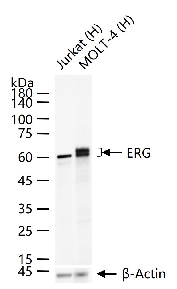
25 ug total protein per lane of various lysates (see on figure) probed with ERG monoclonal antibody, unconjugated (bsm-52324R) at 1:1000 dilution and 4°C overnight incubation. Followed by conjugated secondary antibody incubation at r.t. for 60 min.
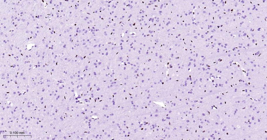
Paraformaldehyde-fixed, paraffin embedded Rat Cerebrum; Antigen retrieval by boiling in sodium citrate buffer (pH6.0) for 15 min; Antibody incubation with ERG Monoclonal Antibody, Unconjugated(bsm-52324R) at 1:200 overnight at 4°C, followed by conjugation to the bs-0295G-HRP and DAB (C-0010) staining.
Paraformaldehyde-fixed, paraffin embedded Mouse Cerebrum; Antigen retrieval by boiling in sodium citrate buffer (pH6.0) for 15 min; Antibody incubation with ERG Monoclonal Antibody, Unconjugated(bsm-52324R) at 1:200 overnight at 4°C, followed by conjugation to the bs-0295G-HRP and DAB (C-0010) staining.
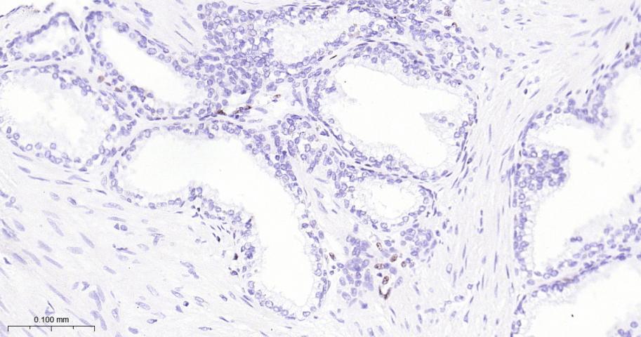
Paraformaldehyde-fixed, paraffin embedded Human Prostate Tumor; Antigen retrieval by boiling in sodium citrate buffer (pH6.0) for 15 min; Antibody incubation with ERG Monoclonal Antibody, Unconjugated(bsm-52324R) at 1:200 overnight at 4°C, followed by conjugation to the bs-0295G-HRP and DAB (C-0010) staining.
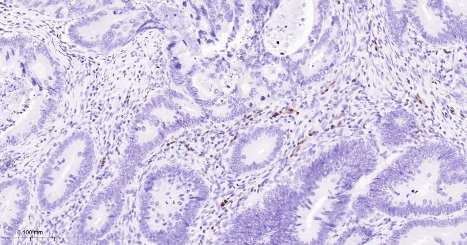
Paraformaldehyde-fixed, paraffin embedded Human Colon Cancer; Antigen retrieval by boiling in sodium citrate buffer (pH6.0) for 15 min; Antibody incubation with ERG Monoclonal Antibody, Unconjugated(bsm-52324R) at 1:200 overnight at 4°C, followed by conjugation to the bs-0295G-HRP and DAB (C-0010) staining.
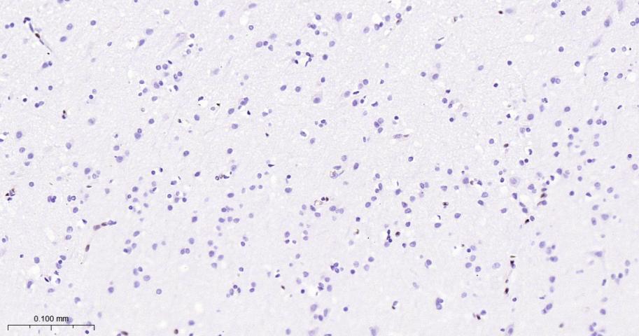
Paraformaldehyde-fixed, paraffin embedded Human Cerebrum; Antigen retrieval by boiling in sodium citrate buffer (pH6.0) for 15 min; Antibody incubation with ERG Monoclonal Antibody, Unconjugated(bsm-52324R) at 1:200 overnight at 4°C, followed by conjugation to the bs-0295G-HRP and DAB (C-0010) staining.
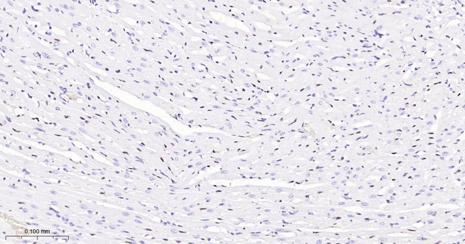
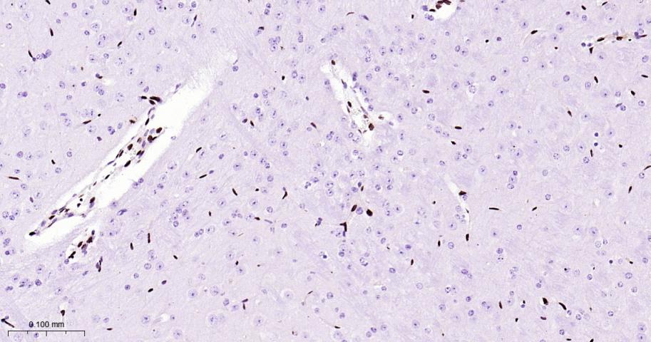
Paraformaldehyde-fixed, paraffin embedded Rat Heart; Antigen retrieval by boiling in sodium citrate buffer (pH6.0) for 15 min; Antibody incubation with ERG Monoclonal Antibody, Unconjugated(bsm-52324R) at 1:200 overnight at 4°C, followed by conjugation to the bs-0295G-HRP and DAB (C-0010) staining.
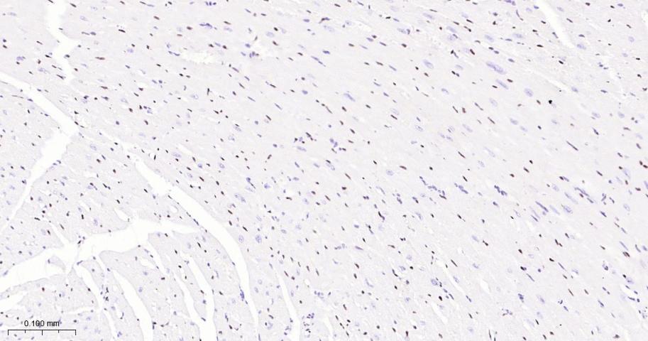
Paraformaldehyde-fixed, paraffin embedded Mouse Heart; Antigen retrieval by boiling in sodium citrate buffer (pH6.0) for 15 min; Antibody incubation with ERG Monoclonal Antibody, Unconjugated(bsm-52324R) at 1:200 overnight at 4°C, followed by conjugation to the bs-0295G-HRP and DAB (C-0010) staining.
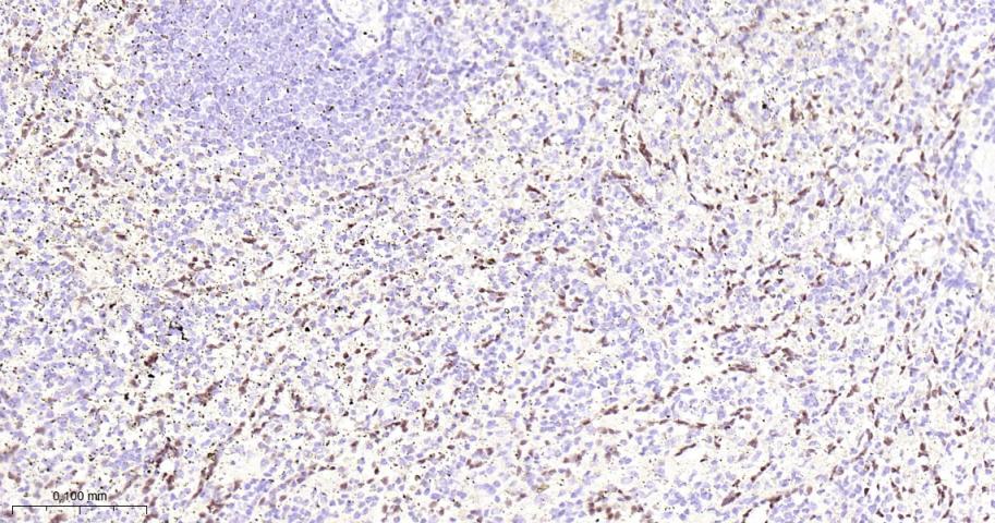
Paraformaldehyde-fixed, paraffin embedded Human Spleen; Antigen retrieval by boiling in sodium citrate buffer (pH6.0) for 15 min; Antibody incubation with ERG Monoclonal Antibody, Unconjugated(bsm-52324R) at 1:200 overnight at 4°C, followed by conjugation to the bs-0295G-HRP and DAB (C-0010) staining.
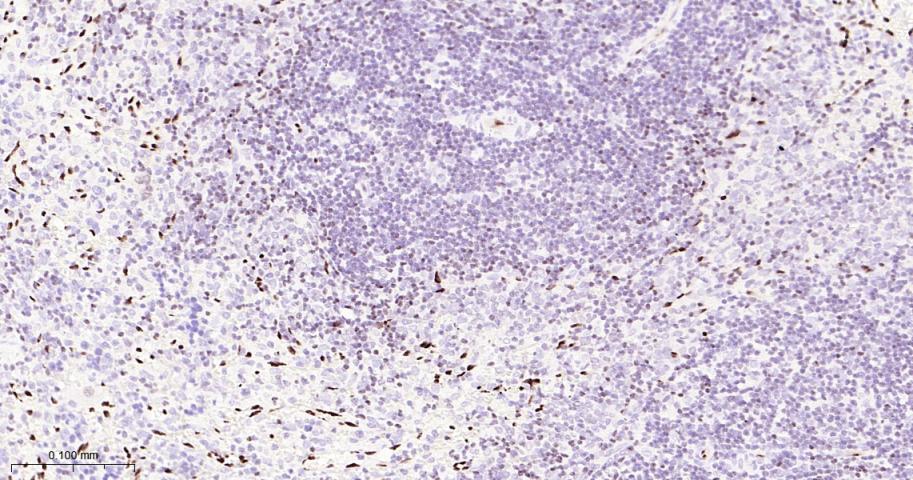
Paraformaldehyde-fixed, paraffin embedded Rat Spleen; Antigen retrieval by boiling in sodium citrate buffer (pH6.0) for 15 min; Antibody incubation with ERG Monoclonal Antibody, Unconjugated(bsm-52324R) at 1:200 overnight at 4°C, followed by conjugation to the bs-0295G-HRP and DAB (C-0010) staining.
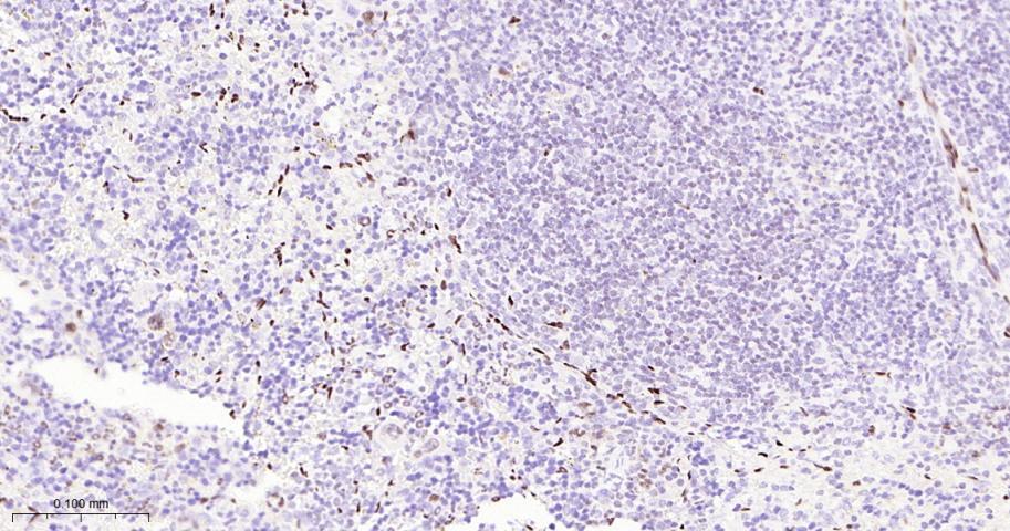
Paraformaldehyde-fixed, paraffin embedded Mouse Spleen; Antigen retrieval by boiling in sodium citrate buffer (pH6.0) for 15 min; Antibody incubation with ERG Monoclonal Antibody, Unconjugated(bsm-52324R) at 1:200 overnight at 4°C, followed by conjugation to the bs-0295G-HRP and DAB (C-0010) staining.
Paraformaldehyde-fixed, paraffin embedded Rat Lung; Antigen retrieval by boiling in sodium citrate buffer (pH6.0) for 15 min; Antibody incubation with ERG Monoclonal Antibody, Unconjugated(bsm-52324R) at 1:200 overnight at 4°C, followed by conjugation to the bs-0295G-HRP and DAB (C-0010) staining.
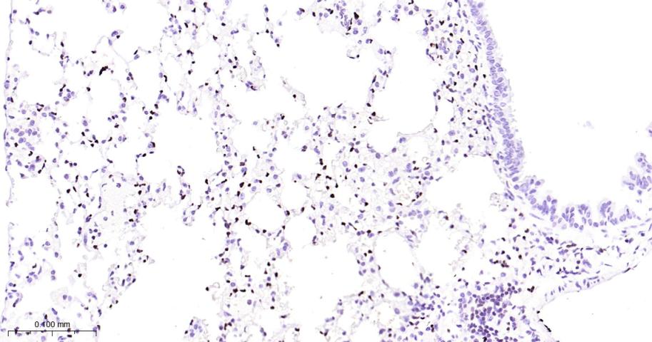
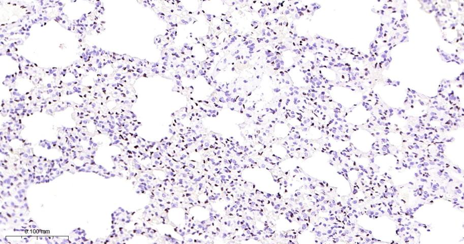
Paraformaldehyde-fixed, paraffin embedded Mouse Lung; Antigen retrieval by boiling in sodium citrate buffer (pH6.0) for 15 min; Antibody incubation with ERG Monoclonal Antibody, Unconjugated(bsm-52324R) at 1:200 overnight at 4°C, followed by conjugation to the bs-0295G-HRP and DAB (C-0010) staining.
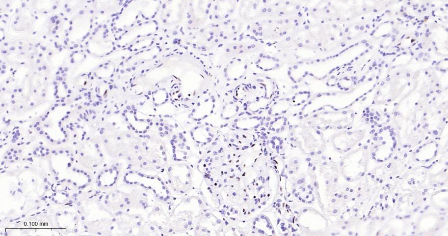
Paraformaldehyde-fixed, paraffin embedded Human Kidney; Antigen retrieval by boiling in sodium citrate buffer (pH6.0) for 15 min; Antibody incubation with ERG Monoclonal Antibody, Unconjugated(bsm-52324R) at 1:200 overnight at 4°C, followed by conjugation to the bs-0295G-HRP and DAB (C-0010) staining.
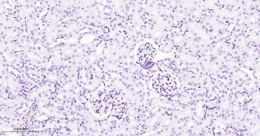
Paraformaldehyde-fixed, paraffin embedded Mouse Kidney; Antigen retrieval by boiling in sodium citrate buffer (pH6.0) for 15 min; Antibody incubation with ERG Monoclonal Antibody, Unconjugated(bsm-52324R) at 1:200 overnight at 4°C, followed by conjugation to the bs-0295G-HRP and DAB (C-0010) staining.
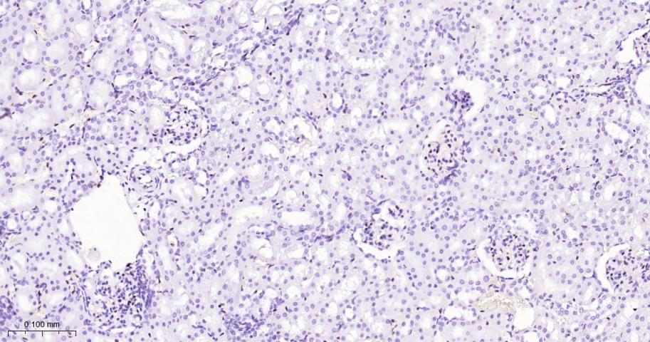
Paraformaldehyde-fixed, paraffin embedded Rat Kidney; Antigen retrieval by boiling in sodium citrate buffer (pH6.0) for 15 min; Antibody incubation with ERG Monoclonal Antibody, Unconjugated(bsm-52324R) at 1:200 overnight at 4°C, followed by conjugation to the bs-0295G-HRP and DAB (C-0010) staining.
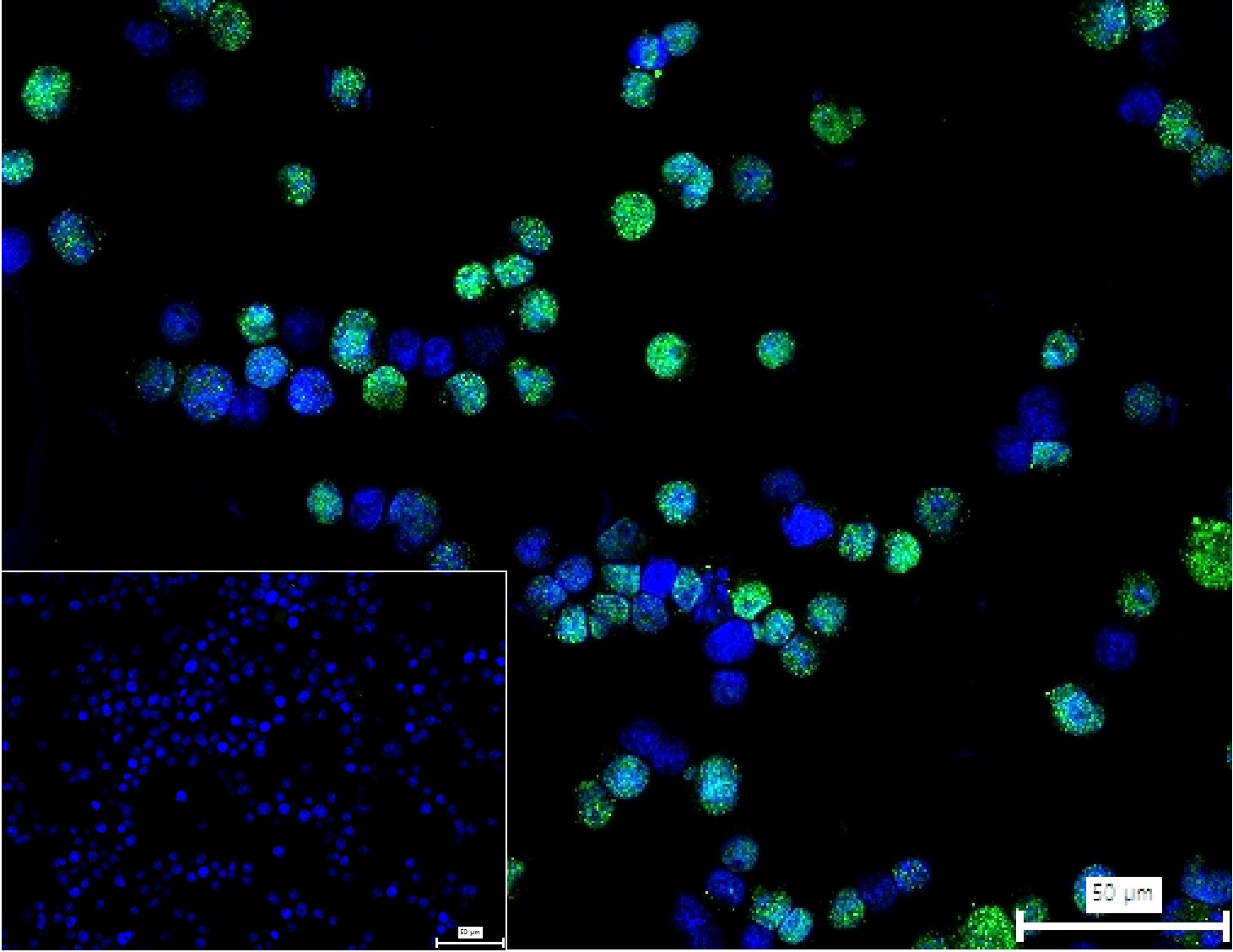
4% Paraformaldehyde-fixed Jurkat (H) cell; Triton X-100 at r.t. for 20 min; Antibody incubation with (ERG) monoclonal Antibody, unconjugated (bsm-52324R) 1:100, 90 min at 37°C; followed by conjugated Goat Anti-Rabbit IgG antibody (green, bs-60295G-BF488) at 37°C for 90 min, DAPI (blue, C02-04002) was used to stain the cell nuclei. PBS instead of the primary antibody was used as the blank control.
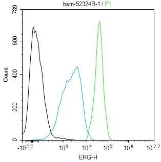
The Jurkat (H) cells were fixed with 4% PFA (10 min at r.t.) and then permeabilized with 90% ice-cold methanol for 20 min at -20℃,the cells then were incubated in 5%BSA to block non-specific protein-protein interactions (30 min at r.t.).Primary Antibody (green):Rabbit Anti-ERG antibody (bsm-52324R,1:100); Secondary Antibody (white blue): Goat anti-Rabbit IgG-BF488(bs-60295G-BF488): 1 μg/test. Blank control (black): PBS. Acquisition of 20,000 events was performed.
|
| 1、抗体溶解方法 | |
| 2、抗体修复方式 | |
| 3、常用试剂的配制 | |
| 4、免疫组化操作步骤 | |
| 5、免疫组化问题解答 | |
| 6、Western Blotting 操作步骤 | |
| 7、Western Blotting 问题解答 | |
| 8、关于肽链的设计 | |
| 9、多肽的溶解与保存 | |
| 10、酶标抗体效价测定程序 | |


