
| 产品编号 | bsm-52774R |
| 英文名称 | CCR7 Recombinant Rabbit mAb |
| 中文名称 | 细胞表面趋化因子受体7重组兔单抗 |
| 别 名 | BLR2; CC-CKR-7; CCR-7; CD197; CDw197; CMKBR7; EBI1; Ebi1h; CCR7_HUMAN; CCR7; C-C CKR-7; Epstein-Barr virus-induced G-protein coupled receptor 1 (EBI1 | EBV-induced G-protein coupled receptor 1); MIP-3 beta receptor; EVI1; CCR7_MOUSE; C-C motif chemokine receptor 7; chemokine (C-C motif) receptor 7 |
| 研究领域 | 免疫学 信号转导 G蛋白偶联受体 G蛋白信号 |
| 抗体来源 | Rabbit |
| 克隆类型 | Recombinant |
| 克 隆 号 | 8C1 |
| 交叉反应 | Human,Mouse,Rat |
| 产品应用 | WB=1:500-2000,Flow-Cyt=1ug/Test,ICC/IF=1:50-200
not yet tested in other applications. optimal dilutions/concentrations should be determined by the end user. |
| 理论分子量 | 42kDa |
| 检测分子量 | 45 |
| 细胞定位 | 细胞膜 |
| 性 状 | Liquid |
| 浓 度 | 1mg/ml |
| 免 疫 原 | A synthesized peptide derived from human CCR7: 1-62 |
| 亚 型 | IgG |
| 纯化方法 | affinity purified by Protein A |
| 缓 冲 液 | 0.01M TBS (pH7.4) with 1% BSA, 0.02% Proclin300 and 50% Glycerol. |
| 保存条件 | Shipped at 4℃. Store at -20℃ for one year. Avoid repeated freeze/thaw cycles. |
| 注意事项 | This product as supplied is intended for research use only, not for use in human, therapeutic or diagnostic applications. |
| PubMed | PubMed |
| 产品介绍 |
The protein encoded by this gene is a member of the G protein-coupled receptor family. This receptor was identified as a gene induced by the Epstein-Barr virus (EBV), and is thought to be a mediator of EBV effects on B lymphocytes. This receptor is expressed in various lymphoid tissues and activates B and T lymphocytes. It has been shown to control the migration of memory T cells to inflamed tissues, as well as stimulate dendritic cell maturation. The chemokine (C-C motif) ligand 19 (CCL19/ECL) has been reported to be a specific ligand of this receptor. [provided by RefSeq, Jul 2008] Function: Receptor for the MIP-3-beta chemokine. Probable mediator of EBV effects on B-lymphocytes or of normal lymphocyte functions. Subcellular Location: Cell membrane; Multi-pass membrane protein. Tissue Specificity: Expressed in various lymphoid tissues and activated B- and T-lymphocytes, strongly up-regulated in B-cells infected with Epstein-Barr virus and T-cells infected with herpesvirus 6 or 7. Similarity: Belongs to the G-protein coupled receptor 1 family. SWISS: P32248 Gene ID: 1236 Database links: Entrez Gene: 1236 Human Entrez Gene: 12775 Mouse Omim: 600242 Human SwissProt: P32248 Human SwissProt: P47774 Mouse Unigene: 370036 Human Unigene: 2932 Mouse Unigene: 229736 Rat 趋化因子是当今细胞因子研究领域的热点之一,它参与多种免疫及炎症反应,在感染、肿瘤的生长与转移、组织修复及创伤愈合等病理生理过程中发挥重要作用.CCR7在肿瘤的生长与转移方面起到一定的作用. |
| 产品图片 |
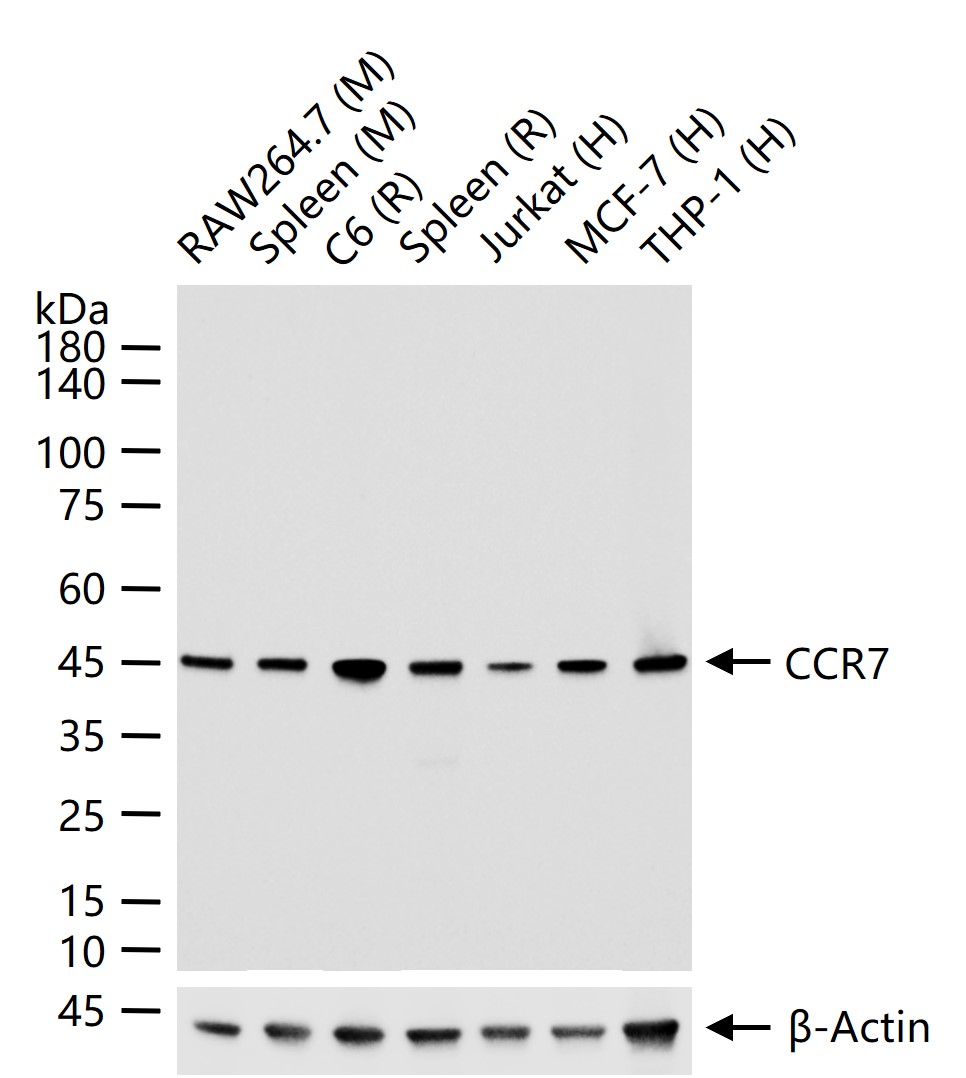
25 ug total protein per lane of various lysates (see on figure) probed with CCR7 monoclonal antibody, unconjugated (bsm-52774R) at 1:1000 dilution and 4°C overnight incubation. Followed by conjugated secondary antibody incubation at r.t. for 60 min.
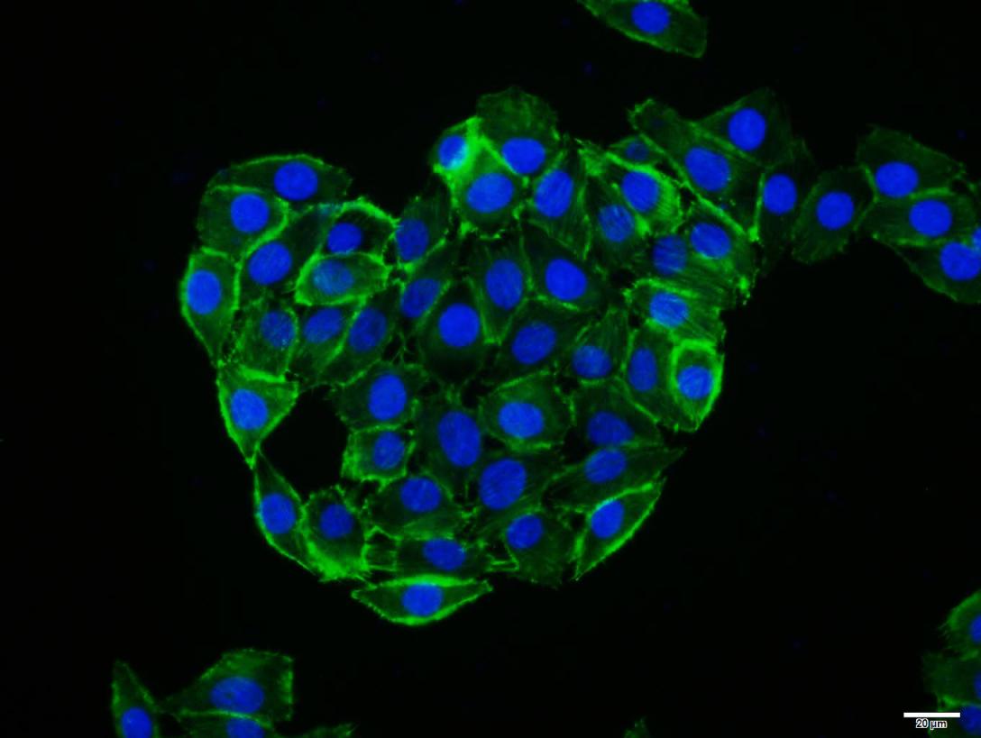
4% Paraformaldehyde-fixed MCF-7 (H) cell; Triton X-100 at r.t. for 20 min; Antibody incubation with (CCR7) monoclonal Antibody, unconjugated (bsm-52774R) 1:100, 90 min at 37°C; followed by conjugated Goat Anti-Rabbit IgG antibody (green, bs-60295G-BF488) at 37°C for 90 min, DAPI (blue, C02-04002) was used to stain the cell nuclei.
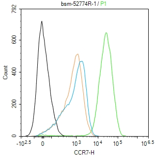
Blank control(black line):Jurkat.
Primary Antibody (green line): Rabbit Anti-CCR7 antibody (bsm-52774R)
Dilution:1ug/Test;
Secondary Antibody(white blue line): Goat anti-Rabit IgG-AF488
Dilution: 0.5ug/Test.
Isotype control(orange line): Normal Rabbit IgG
Protocol
The cells were fixed with 4% PFA (10min at room temperature)and then permeabilized with 90% ice-cold methanol for 20 min at -20℃, The cells were then incubated in 5%BSA to block non-specific protein-protein interactions for 30 min at room temperature .Cells stained with Primary Antibody for 30 min at room temperature. The secondary antibody used for 40 min at room temperature. Acquisition of 20,000 events was performed.
|
| 1、抗体溶解方法 | |
| 2、抗体修复方式 | |
| 3、常用试剂的配制 | |
| 4、免疫组化操作步骤 | |
| 5、免疫组化问题解答 | |
| 6、Western Blotting 操作步骤 | |
| 7、Western Blotting 问题解答 | |
| 8、关于肽链的设计 | |
| 9、多肽的溶解与保存 | |
| 10、酶标抗体效价测定程序 | |


