
| 产品编号 | bsm-52902R |
| 英文名称 | EDG1 Recombinant Rabbit mAb |
| 中文名称 | 内皮细胞分化鞘脂G蛋白偶联受体1重组兔单抗 |
| 别 名 | EDG1; S1P1; CD363; CHEDG1; D1S3362; ECGF1; EDG-1; Lpb1; S1p; S1PR1_BOVIN; S1PR1; S1P receptor 1; Endothelial differentiation G-protein coupled receptor 1; Sphingosine 1-phosphate receptor Edg-1 (S1P receptor Edg-1); S1PR1_HUMAN; S1PR1_MOUSE; Lysophospholi |
| 研究领域 | 细胞生物 免疫学 细胞凋亡 细胞粘附分子 G蛋白偶联受体 内皮细胞 细胞骨架 |
| 抗体来源 | Rabbit |
| 克隆类型 | Recombinant |
| 克 隆 号 | |
| 交叉反应 | Human,Mouse,Rat |
| 产品应用 | WB=1:500-2000
not yet tested in other applications. optimal dilutions/concentrations should be determined by the end user. |
| 理论分子量 | 44 kDa |
| 检测分子量 | 44 |
| 细胞定位 | 细胞膜 |
| 性 状 | Liquid |
| 浓 度 | 1mg/ml |
| 免 疫 原 | KLH conjugated synthetic peptide derived from human EDG1: 2-51/382 |
| 亚 型 | IgG |
| 纯化方法 | affinity purified by Protein A |
| 缓 冲 液 | 0.01M TBS (pH7.4) with 1% BSA, 0.02% Proclin300 and 50% Glycerol. |
| 保存条件 | Shipped at 4℃. Store at -20℃ for one year. Avoid repeated freeze/thaw cycles. |
| 注意事项 | This product as supplied is intended for research use only, not for use in human, therapeutic or diagnostic applications. |
| PubMed | PubMed |
| 产品介绍 |
Sphingosine-1-phosphate receptor 1 (S1P receptor 1 or S1P1), also known as endothelial differentiation gene 1 (EDG1) is a protein that in humans is encoded by the S1PR1 gene. S1PR1 is a G-protein-coupled receptor which binds the bioactive signaling molecule sphingosine 1-phosphate (S1P). S1PR1 belongs to a sphingosine-1-phosphate receptor subfamily comprising five members (S1PR1-5). S1PR1 was originally identified as an abundant transcript in endothelial cells and it has an important role in regulating endothelial cell cytoskeletal structure, migration, capillary-like network formation and vascular maturation. In addition, S1PR1 signaling is important in the regulation of lymphocyte maturation, migration and trafficking. Function: Receptor for the lysosphingolipid sphingosine 1-phosphate (S1P). S1P is a bioactive lysophospholipid that elicits diverse physiological effect on most types of cells and tissues. This inducible epithelial cell G-protein-coupled receptor may be involved in the processes that regulate the differentiation of endothelial cells. Seems to be coupled to the G(i) subclass of heteromeric G proteins. Subcellular Location: Cell membrane; Multi-pass membrane protein. Tissue Specificity: Endothelial cells, and to a lesser extent, in vascular smooth muscle cells, fibroblasts, melanocytes, and cells of epithelioid origin. Post-translational modifications: S1P-induced endothelial cell migration requires the PKB/AKT1-mediated phosphorylation of the third intracellular loop at the Thr-236 residue. Similarity: Belongs to the G-protein coupled receptor 1 family. SWISS: P21453 Gene ID: 1901 Database links: Entrez Gene: 100050049 Horse Entrez Gene: 1901 Human Entrez Gene: 13609 Mouse Omim: 601974 Human SwissProt: P21453 Human SwissProt: O08530 Mouse Unigene: 154210 Human Unigene: 982 Mouse Unigene: 109455 Rat 研究发现,S1P1与VEGF肩并肩相互协作,从而促进血管生长。VEGF是很多不同抗癌药物的作用靶标。血管新生在很多疾病是异常的。靶向S1P1和VEGF可能要比只使用VEGF抑制剂更加有效地治疗疾病;S1P1是一种关键性的血管新生反应性基因。研究人员证实当新的血管网络形成时,由此产生的血液流动激活血管内皮细胞表面上的S1P1,并把信号传递到这些细胞内部从而让新形成的血管网络稳定化。因此阻断S1P1可能有助于切断给癌性肿瘤提供资源的血液供应。 |
| 产品图片 |
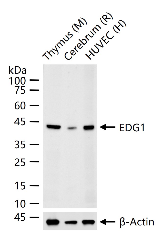
25 ug total protein per lane of various lysates (see on figure) probed with EDG1 monoclonal antibody, unconjugated (bsm-52902R) at 1:1000 dilution and 4°C overnight incubation. Followed by conjugated secondary antibody incubation at r.t. for 60 min.
|
| 1、抗体溶解方法 | |
| 2、抗体修复方式 | |
| 3、常用试剂的配制 | |
| 4、免疫组化操作步骤 | |
| 5、免疫组化问题解答 | |
| 6、Western Blotting 操作步骤 | |
| 7、Western Blotting 问题解答 | |
| 8、关于肽链的设计 | |
| 9、多肽的溶解与保存 | |
| 10、酶标抗体效价测定程序 | |


