
| 产品编号 | bsm-54727R |
| 英文名称 | CHMP2B Recombinant Rabbit mAb |
| 中文名称 | 染色质修饰蛋白2B重组兔单抗 |
| 别 名 | ALS17; CHMP2.5; DMT1; FTDALS7; VPS2-2; VPS2B; 1190006E07Rik; CHM2B_HUMAN; CHMP2B; Chromatin-modifying protein 2b (CHMP2b); Vacuolar protein sorting-associated protein 2-2 (Vps2-2 | hVps2-2); CHM2B_MOUSE; |
| 研究领域 | 肿瘤 细胞生物 神经生物学 信号转导 |
| 抗体来源 | Rabbit |
| 克隆类型 | Recombinant |
| 克 隆 号 | 6F10 |
| 交叉反应 | Human,Mouse,Rat |
| 产品应用 | WB=1:500-2000,Flow-Cyt=1:50-100,IP=1:20-50
not yet tested in other applications. optimal dilutions/concentrations should be determined by the end user. |
| 理论分子量 | 24 kDa |
| 检测分子量 | 28 |
| 细胞定位 | 细胞浆 |
| 性 状 | Liquid |
| 浓 度 | 1mg/ml |
| 免 疫 原 | Recombinant protein within C-terminal Human CHMP2B. |
| 亚 型 | IgG |
| 纯化方法 | affinity purified by Protein A |
| 缓 冲 液 | 0.01M TBS (pH7.4) with 1% BSA, 0.02% Proclin300 and 50% Glycerol. |
| 保存条件 | Shipped at 4℃. Store at -20℃ for one year. Avoid repeated freeze/thaw cycles. |
| 注意事项 | This product as supplied is intended for research use only, not for use in human, therapeutic or diagnostic applications. |
| PubMed | PubMed |
| 产品介绍 |
The charged multivesicular body proteins, commonly designated CHMPs, belong to the vacuolar sorting protein family and function as chromatin-modifying proteins. CHMP1-6 are all components of ESCRT (endosomal sorting complex required for transport) I, II or III complexes. These complexes are crucial for sorting endosomal articles into multivesicular bodies (MVBs), and are also required for the formation of these bodies. CHMP2B, also known as CHMP2.5 or vacuolar protein-sorting-associated protein 2-2, is a 213 amino acid cytosolic protein. Widely expressed in brain, heart, skeletal muscle, small intestine, pancreas, lung, placenta and leukocytes, CHMP2B associates directly with CHMP2A and vps4 for the disassembly of the ESCRT-III complex. Defects in the gene encoding CHMP2B have been shown to cause chromosome 3-linked frontotemporal dementia (FTD3). Function: Probable core component of the endosomal sorting required for transport complex III (ESCRT-III) which is involved in multivesicular bodies (MVBs) formation and sorting of endosomal cargo proteins into MVBs. MVBs contain intraluminal vesicles (ILVs) that are generated by invagination and scission from the limiting membrane of the endosome and mostly are delivered to lysosomes enabling degradation of membrane proteins, such as stimulated growth factor receptors, lysosomal enzymes and lipids. The MVB pathway appears to require the sequential function of ESCRT-O, -I,-II and -III complexes. ESCRT-III proteins mostly dissociate from the invaginating membrane before the ILV is released. The ESCRT machinery also functions in topologically equivalent membrane fission events, such as the terminal stages of cytokinesis and the budding of enveloped viruses (HIV-1 and other lentiviruses). ESCRT-III proteins are believed to mediate the necessary vesicle extrusion and/or membrane fission activities, possibly in conjunction with the AAA ATPase VPS4. Subunit: Probable core component of the endosomal sorting required for transport complex III (ESCRT-III). ESCRT-III components are thought to multimerize to form a flat lattice on the perimeter membrane of the endosome. Several assembly forms of ESCRT-III may exist that interact and act sequentally. Interacts with CHMP2A. Interacts with VPS4A. Interacts with VPS4B; the interaction is direct. Subcellular Location: Cytoplasm Tissue Specificity: Widely expressed. Expressed in brain, heart, skeletal muscle, spleen, kidney, liver, small intestine, pancreas, lung, placenta and leukocytes. In brain, it is expressed in cerebellum, cerebral cortex, medulla, spinal chord, occipital lobe, frontal lobe, temporal lobe and putamen. DISEASE: Defects in CHMP2B are the cause of frontotemporal dementia, chromosome 3-linked (FTD3) [MIM:600795]. FTD3 is characterized by an onset of dementia in the late 50's initially characterized by behavioral and personality changes including apathy, restlessness, disinhibition and hyperorality, progressing to stereotyped behaviors, non-fluent aphasia, mutism and dystonia, with a marked lack of insight. The brains of individuals with FTD3 have no distinctive neuropathological features. They show global cortical and central atrophy, but no beta-amyloid deposits. Similarity: Belongs to the SNF7 family. SWISS: Q9UQN3 Gene ID: 25978 Database links: Entrez Gene: 25978 Human Entrez Gene: 68942 Mouse Omim: 609512 Human SwissProt: Q9UQN3 Human SwissProt: Q8BJF9 Mouse Unigene: 476930 Human Unigene: 432944 Mouse |
| 产品图片 |
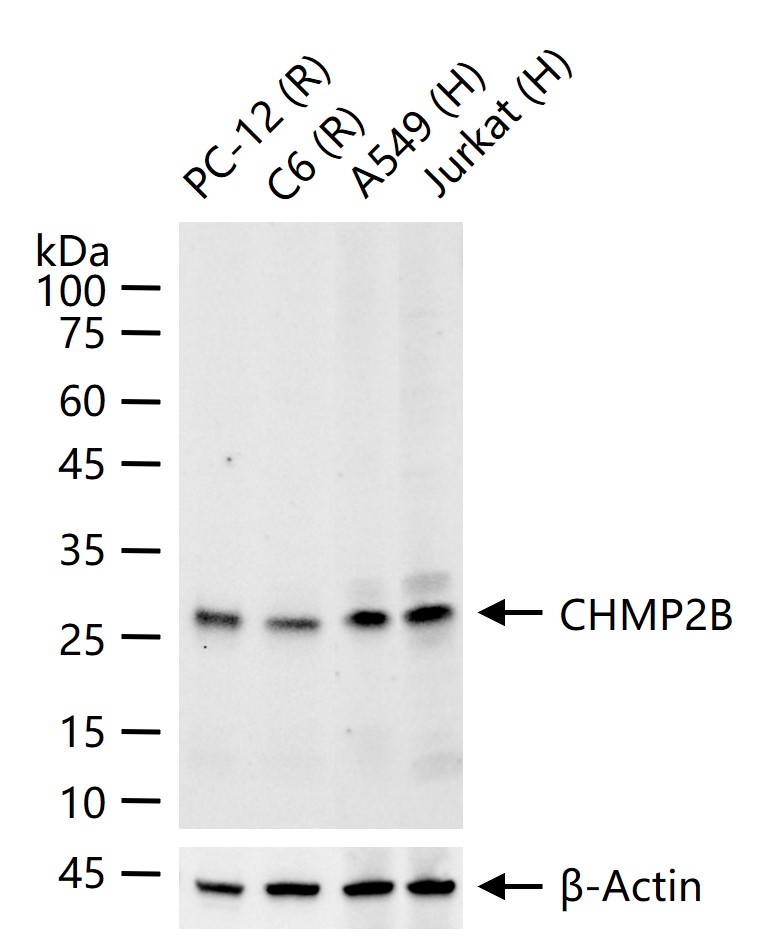
25 ug total protein per lane of various lysates (see on figure) probed with CHMP2B monoclonal antibody, unconjugated (bsm-54727R) at 1:1000 dilution and 4°C overnight incubation. Followed by conjugated secondary antibody incubation at r.t. for 60 min.
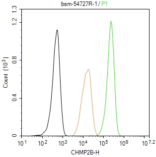
The Hela (H) cells were fixed with 4% PFA (10 min at r.t.) and then permeabilized with 90% ice-cold methanol for 20 min at -20℃,the cells then were incubated in 5%BSA to block non-specific protein-protein interactions (30 min at r.t.), followed by secondary antibody incubation for 40 min at room temperature. Primary Antibody (green):Rabbit Anti-CHMP2B antibody (bsm-54727R,1:100); Isotype Control (orange): Rabbit IgG (bs-0295P). Blank control (black): PBS. Acquisition of 20,000 events was performed.
|
| 1、抗体溶解方法 | |
| 2、抗体修复方式 | |
| 3、常用试剂的配制 | |
| 4、免疫组化操作步骤 | |
| 5、免疫组化问题解答 | |
| 6、Western Blotting 操作步骤 | |
| 7、Western Blotting 问题解答 | |
| 8、关于肽链的设计 | |
| 9、多肽的溶解与保存 | |
| 10、酶标抗体效价测定程序 | |


