
| 产品编号 | bsm-52290R |
| 英文名称 | beta Tubulin Recombinant Rabbit mAb |
| 中文名称 | 微管蛋白重组兔单抗 |
| 别 名 | CDCBM6; CSCSC1; M40; OK/SW-cl.56; TUBB1; TUBB5; B130022C14Rik; M(beta)5; Tubb; TBB5_HUMAN; Tubulin beta-5 chain; TBB5_MOUSE; TBB5_RAT; |
 |
Specific References (1) | bsm-52290R has been referenced in 1 publications.
[IF=5.139] Yingying Che. et al. Splicing factor SRSF3 promotes the progression of cervical cancer through regulating DDX5. MOL CARCINOGEN. 2022 Oct;: WB ; Human.
|
| 研究领域 | 细胞生物 免疫学 神经生物学 细胞骨架 |
| 抗体来源 | Rabbit |
| 克隆类型 | Recombinant |
| 克 隆 号 | 2F11 |
| 交叉反应 | Human,Mouse,Rat |
| 产品应用 | WB=1:2000-5000,IHC-P=1:50-200,IHC-F=1:50-200,IF=1:50-200
not yet tested in other applications. optimal dilutions/concentrations should be determined by the end user. |
| 理论分子量 | 50 kDa |
| 检测分子量 | 50 |
| 细胞定位 | 细胞浆 |
| 性 状 | Liquid |
| 浓 度 | 1mg/ml |
| 免 疫 原 | A synthesized peptide derived from human beta Tubulin: 400-444 |
| 亚 型 | IgG |
| 纯化方法 | affinity purified by Protein A |
| 缓 冲 液 | 0.01M TBS (pH7.4) with 1% BSA, 0.02% Proclin300 and 50% Glycerol. |
| 保存条件 | Shipped at 4℃. Store at -20℃ for one year. Avoid repeated freeze/thaw cycles. |
| 注意事项 | This product as supplied is intended for research use only, not for use in human, therapeutic or diagnostic applications. |
| PubMed | PubMed |
| 产品介绍 |
Microtubules are constituent parts of the mitotic apparatus, cilia, flagella, and elements of the cytoskeleton. They consist principally of 2 soluble proteins, alpha- and beta-tubulin, each of about 55,000 kDa. Antibodies against beta Tubulin are useful as loading controls for Western Blotting. However it should be noted that levels of beta Tubulin may not be stable in certain cells. For example, expression of tubulin in adipose tissue is very low (Spiegelman and Farmer, Cell, 1982, 29(1):53-60) and therefore beta Tubulin should not be used as loading control for these tissues. SWISS: P07437 Gene ID: 203068 |
| 产品图片 |
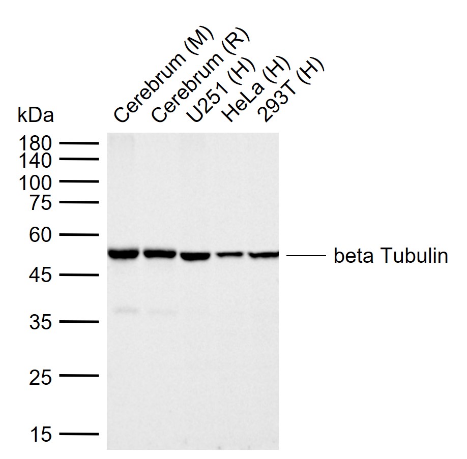
Sample:
Lane 1: Mouse Cerebrum tissue lysates
Lane 2: Rat Cerebrum tissue lysates
Lane 3: Human U251 cell lysates
Lane 4: Human HeLa cell lysates
Lane 5: Human 293T cell lysates
Primary: Anti-beta Tubulin (bsm-52290R) at 1/20000 dilution
Secondary: IRDye800CW Goat Anti-Rabbit IgG at 1/20000 dilution
Predicted band size: 50 kDa
Observed band size: 50 kDa
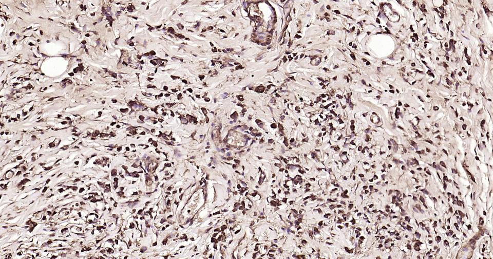
Paraformaldehyde-fixed, paraffin embedded Human Breast Cancer; Antigen retrieval by boiling in sodium citrate buffer (pH6.0) for 15 min; Antibody incubation with beta Tubulin Monoclonal Antibody, Unconjugated(bsm-52290R) at 1:200 overnight at 4°C, followed by conjugation to the SP Kit(Rabbit, SP-0023) and DAB (C-0010) staining.
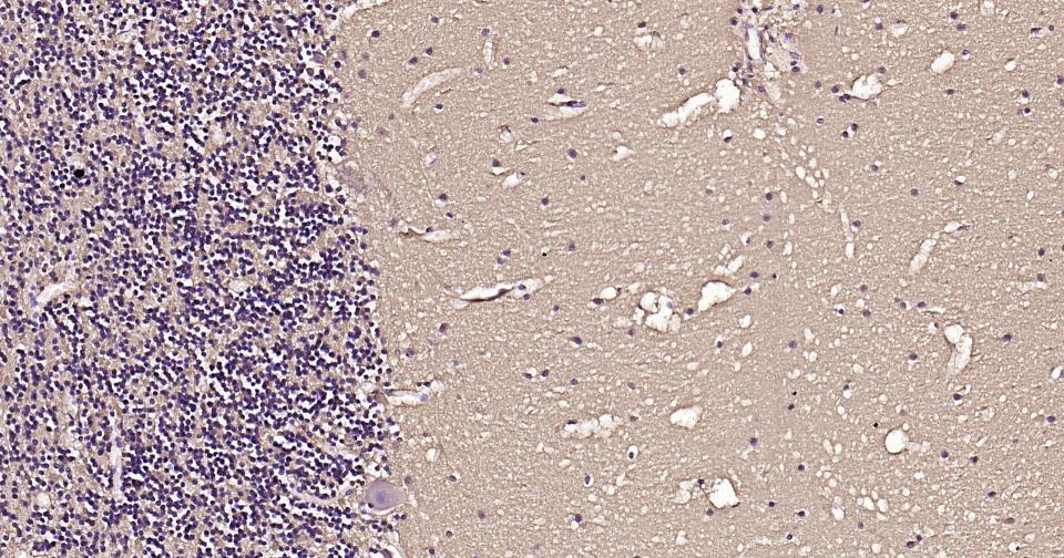
Paraformaldehyde-fixed, paraffin embedded Human Cerebellum; Antigen retrieval by boiling in sodium citrate buffer (pH6.0) for 15 min; Antibody incubation with beta Tubulin Monoclonal Antibody, Unconjugated(bsm-52290R) at 1:200 overnight at 4°C, followed by conjugation to the SP Kit(Rabbit, SP-0023) and DAB (C-0010) staining.
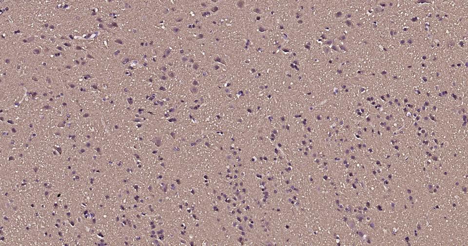
Paraformaldehyde-fixed, paraffin embedded Human Cerebrum; Antigen retrieval by boiling in sodium citrate buffer (pH6.0) for 15 min; Antibody incubation with beta Tubulin Monoclonal Antibody, Unconjugated(bsm-52290R) at 1:200 overnight at 4°C, followed by conjugation to the SP Kit(Rabbit, SP-0023) and DAB (C-0010) staining.
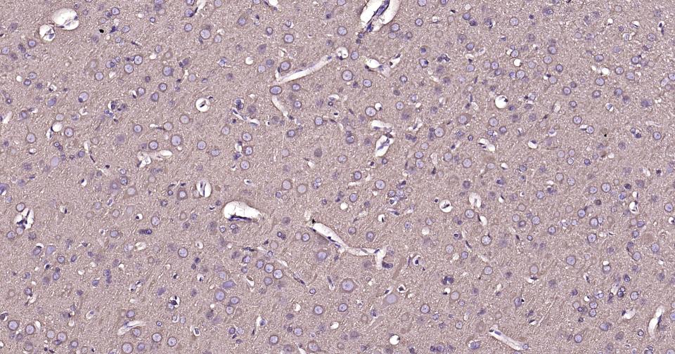
Paraformaldehyde-fixed, paraffin embedded Rat Cerebrum; Antigen retrieval by boiling in sodium citrate buffer (pH6.0) for 15 min; Antibody incubation with beta Tubulin Monoclonal Antibody, Unconjugated(bsm-52290R) at 1:200 overnight at 4°C, followed by conjugation to the SP Kit(Rabbit, SP-0023) and DAB (C-0010) staining.
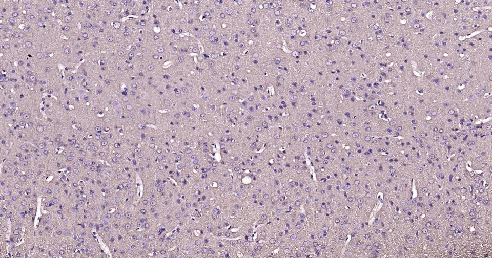
Paraformaldehyde-fixed, paraffin embedded Mouse Cerebrum; Antigen retrieval by boiling in sodium citrate buffer (pH6.0) for 15 min; Antibody incubation with beta Tubulin Monoclonal Antibody, Unconjugated(bsm-52290R) at 1:200 overnight at 4°C, followed by conjugation to the SP Kit(Rabbit, SP-0023) and DAB (C-0010) staining.

Paraformaldehyde-fixed, paraffin embedded Rat Testicles; Antigen retrieval by boiling in sodium citrate buffer (pH6.0) for 15 min; Antibody incubation with beta Tubulin Monoclonal Antibody, Unconjugated(bsm-52290R) at 1:200 overnight at 4°C, followed by conjugation to the SP Kit(Rabbit, SP-0023) and DAB (C-0010) staining.
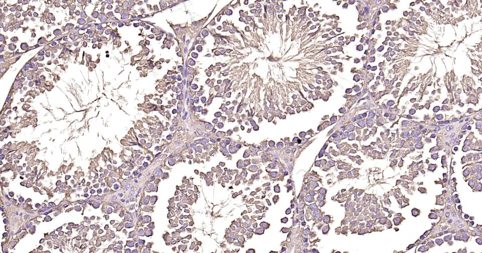
Paraformaldehyde-fixed, paraffin embedded Mouse Testicles; Antigen retrieval by boiling in sodium citrate buffer (pH6.0) for 15 min; Antibody incubation with beta Tubulin Monoclonal Antibody, Unconjugated(bsm-52290R) at 1:200 overnight at 4°C, followed by conjugation to the SP Kit(Rabbit, SP-0023) and DAB (C-0010) staining.
|
| 1、抗体溶解方法 | |
| 2、抗体修复方式 | |
| 3、常用试剂的配制 | |
| 4、免疫组化操作步骤 | |
| 5、免疫组化问题解答 | |
| 6、Western Blotting 操作步骤 | |
| 7、Western Blotting 问题解答 | |
| 8、关于肽链的设计 | |
| 9、多肽的溶解与保存 | |
| 10、酶标抗体效价测定程序 | |


