

| 产品编号 | bs-42054R |
| 英文名称 | CD36 Rabbit pAb |
| 中文名称 | CD36抗体 |
| 别 名 | BDPLT10; CHDS7; FAT; GP3B; GP4; GPIV; PASIV; SCARB3; GPIIIB; IV; PAS; PAS IV; PAS-4; CD36_HUMAN; CD36; Fatty acid translocase (FAT); Glycoprotein IIIb (GPIIIB); Leukocyte differentiation antigen CD36; Platelet collagen receptor; Platelet glycoprotein IV (GPIV); Thrombospondin receptor; CD36_MOUSE; CD36_RAT; Adipocyte membrane protein; Fatty acid translocase; Fatty acid transport protein; |
| 抗体来源 | Rabbit |
| 克隆类型 | Polyclonal |
| 克 隆 号 | |
| 交叉反应 | Mouse |
| 产品应用 | WB=1:500-2000,IHC-P=1:100-500,IHC-F=1:100-500,IF=1:100-500
not yet tested in other applications. optimal dilutions/concentrations should be determined by the end user. |
| 理论分子量 | 52 kDa |
| 检测分子量 | 75-85 |
| 细胞定位 | 细胞膜 |
| 性 状 | Liquid |
| 浓 度 | 1mg/ml |
| 免 疫 原 | Recombinant mouse CD36 protein: 30-439/472 <Extracellular> |
| 亚 型 | IgG |
| 纯化方法 | affinity purified by Protein A |
| 缓 冲 液 | 0.01M TBS (pH7.4) with 1% BSA, 0.02% Proclin300 and 50% Glycerol. |
| 保存条件 | Shipped at 4℃. Store at -20℃ for one year. Avoid repeated freeze/thaw cycles. |
| 注意事项 | This product as supplied is intended for research use only, not for use in human, therapeutic or diagnostic applications. |
| PubMed | PubMed |
| 产品介绍 |
The protein encoded by this gene is the fourth major glycoprotein of the platelet surface and serves as a receptor for thrombospondin in platelets and various cell lines. Since thrombospondins are widely distributed proteins involved in a variety of adhesive processes, this protein may have important functions as a cell adhesion molecule. It binds to collagen, thrombospondin, anionic phospholipids and oxidized LDL. It directly mediates cytoadherence of Plasmodium falciparum parasitized erythrocytes and it binds long chain fatty acids and may function in the transport and/or as a regulator of fatty acid transport. Mutations in this gene cause platelet glycoprotein deficiency. Multiple alternatively spliced transcript variants have been found for this gene. [provided by RefSeq, Feb 2014] Function: Seems to have numerous potential physiological functions. Binds to collagen, thrombospondin, anionic phospholipids and oxidized LDL. May function as a cell adhesion molecule. Directly mediates cytoadherence of Plasmodium falciparum parasitized erythrocytes. Binds long chain fatty acids and may function in the transport and/or as a regulator of fatty acid transport. Receptor for thombospondins, THBS1 AND THBS2, mediating their antiangiogenic effects. Subunit: Interacts with THBS1 and THBS2; the interactions mediate the THBS antiangiogenic activity. Subcellular Location: Membrane; Multi-pass membrane protein. Tissue Specificity: Predominant in heart, intestine, spleen, fat, skeletal muscle, lower in testes. Post-translational modifications: N-glycosylated and O-glycosylated with a ratio of 2:1. DISEASE: Defects in CD36 are the cause of platelet glycoprotein IV deficiency (PG4D)[MIM:608404]; also known as CD36 deficiency. Platelet glycoprotein IV deficiency can be divided into 2 subgroups. The type I phenotype is characterized by platelets and monocytes/macrophages exhibiting complete CD36 deficiency. The type II phenotype lacks the surface expression of CD36 in platelets, but expression in monocytes/macrophages is near normal. Genetic variations in CD36 are associated with susceptibility to coronary heart disease type 7 (CHDS7) [MIM:610938]. Similarity: Belongs to the CD36 family. SWISS: Q08857 Gene ID: 12491 Database links: Entrez Gene: 948 Human Entrez Gene: 12491 Mouse Omim: 173510 Human SwissProt: P16671 Human SwissProt: Q08857 Mouse Unigene: 120949 Human Unigene: 18628 Mouse Unigene: 406799 Mouse Unigene: 102418 Rat Unigene: 205840 Rat CD36属一种多功能细胞膜受体,CD36参与长链脂肪酸的吸附和调节细胞凋亡残物的清理和巨噬细胞的吞噬作用, 属于一种抗蛋白酶血小板膜表面糖蛋白。 CD36 广泛在单核细胞、巨噬细胞、内皮层细胞、血小板、红细胞前板、脂肪细胞、肌肉细胞以及乳腺内皮层细胞中有不同的表达。CD36抗原是血小板反应素的配体,两者结合后产生抑制肿瘤血管生成的效应。 |
| 产品图片 |

25 ug total protein per lane of various lysates (see on figure) probed with CD36 polyclonal antibody, unconjugated (bs-42054R) at 1:1000 dilution and 4°C overnight incubation. Followed by conjugated secondary antibody incubation at r.t. for 60 min.
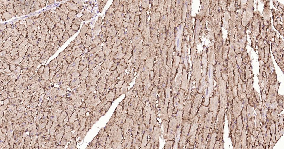
Paraformaldehyde-fixed, paraffin embedded Mouse Heart; Antigen retrieval by boiling in sodium citrate buffer (pH6.0) for 15 min; Antibody incubation with CD36 Polyclonal Antibody, Unconjugated (bs-42054R) at 1:200 overnight at 4°C, followed by conjugation to the SP Kit (Rabbit, SP-0023) and DAB (C-0010) staining.
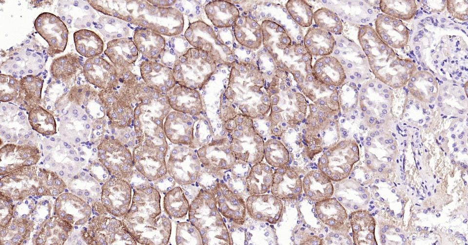
Paraformaldehyde-fixed, paraffin embedded Mouse Kidney; Antigen retrieval by boiling in sodium citrate buffer (pH6.0) for 15 min; Antibody incubation with CD36 Polyclonal Antibody, Unconjugated (bs-42054R) at 1:200 overnight at 4°C, followed by conjugation to the SP Kit (Rabbit, SP-0023) and DAB (C-0010) staining.
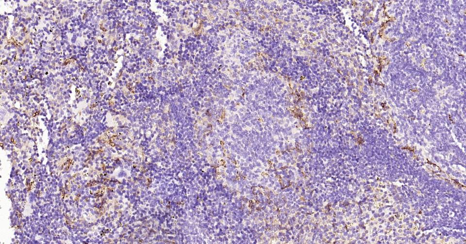
Paraformaldehyde-fixed, paraffin embedded Mouse Spleen; Antigen retrieval by boiling in sodium citrate buffer (pH6.0) for 15 min; Antibody incubation with CD36 Polyclonal Antibody, Unconjugated (bs-42054R) at 1:200 overnight at 4°C, followed by conjugation to the SP Kit (Rabbit, SP-0023) and DAB (C-0010) staining.
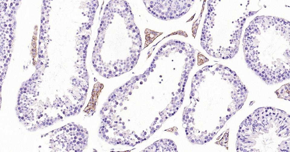
Paraformaldehyde-fixed, paraffin embedded Mouse Testicles; Antigen retrieval by boiling in sodium citrate buffer (pH6.0) for 15 min; Antibody incubation with CD36 Polyclonal Antibody, Unconjugated (bs-42054R) at 1:200 overnight at 4°C, followed by conjugation to the SP Kit (Rabbit, SP-0023) and DAB (C-0010) staining.
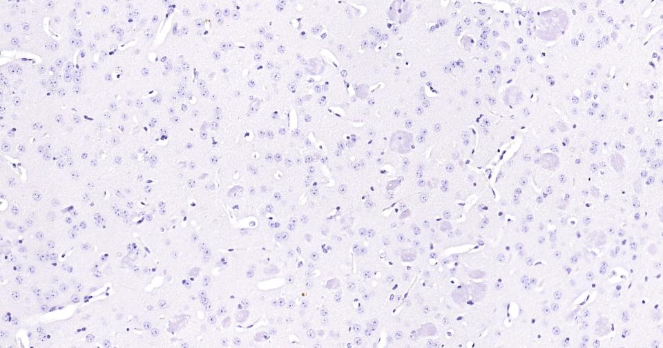
(Negative control) Paraformaldehyde-fixed, paraffin embedded Mouse Cerebrum; Antigen retrieval by boiling in sodium citrate buffer (pH6.0) for 15 min; Antibody incubation with CD36 Polyclonal Antibody, Unconjugated (bs-42054R) at 1:200 overnight at 4°C, followed by conjugation to the SP Kit (Rabbit, SP-0023) and DAB (C-0010) staining.
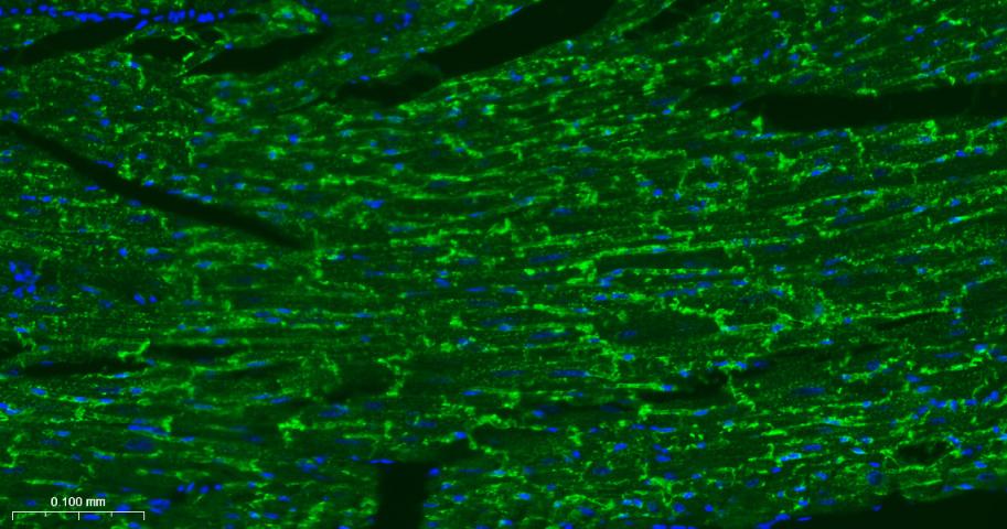
Paraformaldehyde-fixed, paraffin embedded Mouse Heart; Antigen retrieval by boiling in sodium citrate buffer (pH6.0) for 15 min; Antibody incubation with CD36 Polyclonal Antibody, Unconjugated (bs-42054R) at 1:200 overnight at 4°C. Followed by conjugated Goat Anti-Rabbit IgG antibody (green, bs-0295G-BF488), DAPI (blue, C02-04002) was used to stain the cell nuclei.
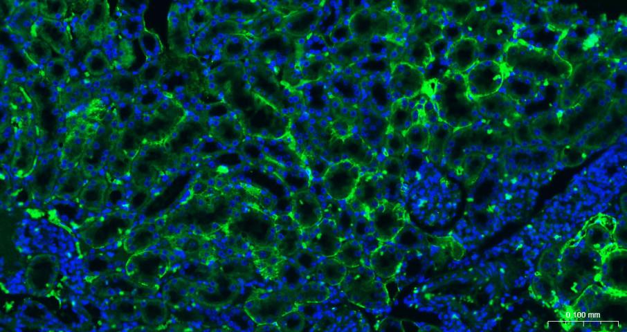
Paraformaldehyde-fixed, paraffin embedded Mouse Kidney; Antigen retrieval by boiling in sodium citrate buffer (pH6.0) for 15 min; Antibody incubation with CD36 Polyclonal Antibody, Unconjugated (bs-42054R) at 1:200 overnight at 4°C. Followed by conjugated Goat Anti-Rabbit IgG antibody (green, bs-0295G-BF488), DAPI (blue, C02-04002) was used to stain the cell nuclei.
|
| 1、抗体溶解方法 | |
| 2、抗体修复方式 | |
| 3、常用试剂的配制 | |
| 4、免疫组化操作步骤 | |
| 5、免疫组化问题解答 | |
| 6、Western Blotting 操作步骤 | |
| 7、Western Blotting 问题解答 | |
| 8、关于肽链的设计 | |
| 9、多肽的溶解与保存 | |
| 10、酶标抗体效价测定程序 | |


