
| 产品编号 | bsm-52259R |
| 英文名称 | ERK1/2 Recombinant Rabbit mAb |
| 中文名称 | 丝裂原活化蛋白激酶1/ERK 1/2重组兔单抗 |
| 别 名 | MK03_HUMAN; MAPK3; MAP kinase 3; MAPK 3; ERT2; Extracellular signal-regulated kinase 1 (ERK-1); Insulin-stimulated MAP2 kinase; MAP kinase isoform p44 (p44-MAPK); Microtubule-associated protein 2 kinase; p44-ERK1; 2.7.11.24; ERK1; PRKM3; MK01_HUMAN; MAPK1 |
 |
Specific References (5) | bsm-52259R has been referenced in 5 publications.
[IF=7.675] Honghong Zhan. et al. Oxybaphus himalaicus Mitigates Lipopolysaccharide-Induced Acute Kidney Injury by Inhibiting TLR4/MD2 Complex Formation. ANTIOXIDANTS-BASEL. 2022 Dec;11(12):2307 WB ; Mouse.
[IF=4.411] Yudan Zhao. et al. COX-2 is required to mediate crosstalk of ROS-dependent activation of MAPK/NF-κB signaling with pro-inflammatory response and defense-related NO enhancement during challenge of macrophage-like cell line with Giardia duodenalis. PLOS NEGLECT TROP D. 2022 Apr;16(4):e0010402 WB ; Mouse.
[IF=4.319] Jia Li. et al. Study of the Mechanism of Antiemetic Effect of Lavandula angustifolia Mill. Essential Oil Based on Ca2+/CaMKII/ERK1/2 Pathway. DRUG DES DEV THER. 2022 Jul;16:2407-2422 IHC ; Rat.
[IF=2.65] Yiming Bi. et al. β-Sitosterol Suppresses LPS-Induced Cytokine Production in Human Umbilical Vein Endothelial Cells via MAPKs and NF-κB Signaling Pathway. EVID-BASED COMPL ALT. 2023 Jan 03;2023:9241090 WB ; Human.
[IF=0] Xing-Peng Di. et al. YAP/Smad3 promotes pathological extracellular matrix microenviroment-induced bladder smooth muscle proliferation in bladder fibrosis progression. MedComm. 2022 Sep;3(4):e169 WB ; Human.
|
| 研究领域 | 肿瘤 细胞生物 神经生物学 干细胞 细胞凋亡 转录调节因子 激酶和磷酸酶 细胞骨架 |
| 抗体来源 | Rabbit |
| 克隆类型 | Recombinant |
| 克 隆 号 | 3A12 |
| 交叉反应 | Human,Mouse,Rat |
| 产品应用 | WB=1:1000-5000,IHC-P=1:200-800,IHC-F=1:200-800,IF=1:200-800,Flow-Cyt=1ug/Test,ICC/IF=1:100-500
not yet tested in other applications. optimal dilutions/concentrations should be determined by the end user. |
| 理论分子量 | 42/44 kDa |
| 检测分子量 | 42/44 |
| 细胞定位 | 细胞核 细胞浆 细胞膜 细胞外基质 |
| 性 状 | Liquid |
| 浓 度 | 1mg/ml |
| 免 疫 原 | KLH conjugated synthetic peptide derived from human ERK1/2 |
| 亚 型 | IgG |
| 纯化方法 | affinity purified by Protein A |
| 缓 冲 液 | 0.01M TBS (pH7.4) with 1% BSA, 0.02% Proclin300 and 50% Glycerol. |
| 保存条件 | Store at -20℃ for one year. Avoid repeated freeze/thaw cycles. |
| 注意事项 | This product as supplied is intended for research use only, not for use in human, therapeutic or diagnostic applications. |
| PubMed | PubMed |
| 产品介绍 |
The protein encoded by this gene is a member of the MAPkinase family. MAP kinases, also known as extracellularsignal-regulated kinases (ERKs), act in a signaling cascade thatregulates various cellular processes such as proliferation,differentiation, and cell cycle progression in response to avariety of extracellular signals. This kinase is activated byupstream kinases, resulting in its translocation to the nucleuswhere it phosphorylates nuclear targets. Alternatively splicedtranscript variants encoding different protein isoforms have beendescribed. [provided by RefSeq, Jul 2008]. Function: Serine/threonine kinase which acts as an essentialcomponent of the MAP kinase signal transduction pathway. MAPK1/ERK2and MAPK3/ERK1 are the 2 MAPKs which play an important role in theMAPK/ERK cascade. They participate also in a signaling cascadeinitiated by activated KIT and KITLG/SCF. Depending on the cellularcontext, the MAPK/ERK cascade mediates diverse biological functionssuch as cell growth, adhesion, survival and differentiation throughthe regulation of transcription, translation, cytoskeletalrearrangements. The MAPK/ERK cascade plays also a role ininitiation and regulation of meiosis, mitosis, and postmitoticfunctions in differentiated cells by phosphorylating a number oftranscription factors. About 160 substrates have already beendiscovered for ERKs. Many of these substrates are localized in thenucleus, and seem to participate in the regulation of transcriptionupon stimulation. However, other substrates are found in thecytosol as well as in other cellular organelles, and those areresponsible for processes such as translation, mitosis andapoptosis. Moreover, the MAPK/ERK cascade is also involved in theregulation of the endosomal dynamics, including lysosome processingand endosome cycling through the perinuclear recycling compartment(PNRC); as well as in the fragmentation of the Golgi apparatusduring mitosis. The substrates include transcription factors (suchas ATF2, BCL6, ELK1, ERF, FOS, HSF4 or SPZ1), cytoskeletal elements(such as CANX, CTTN, GJA1, MAP2, MAPT, PXN, SORBS3 or STMN1),regulators of apoptosis (such as BAD, BTG2, CASP9, DAPK1, IER3,MCL1 or PPARG), regulators of translation (such as EIF4EBP1) and avariety of other signaling-related molecules (like ARHGEF2, DCC,FRS2 or GRB10). Protein kinases (such as RAF1, RPS6KA1/RSK1,RPS6KA3/RSK2, RPS6KA2/RSK3, RPS6KA6/RSK4, SYK, MKNK1/MNK1,MKNK2/MNK2, RPS6KA5/MSK1, RPS6KA4/MSK2, MAPKAPK3 or MAPKAPK5) andphosphatases (such as DUSP1, DUSP4, DUSP6 or DUSP16) are othersubstrates which enable the propagation the MAPK/ERK signal toadditional cytosolic and nuclear targets, thereby extending thespecificity of the cascade. Mediates phosphorylation of TPR inrespons to EGF stimulation. May play a role in the spindle assemblycheckpoint. Phosphorylates PML and promotes its interaction withPIN1, leading to PML degradation (By similarity). Acts as a transcriptional repressor. Binds to a[GC]AAA[GC] consensus sequence. Repress the expression ofinterferon gamma-induced genes. Seems to bind to the promoter ofCCL5, DMP1, IFIH1, IFITM1, IRF7, IRF9, LAMP3, OAS1, OAS2, OAS3 andSTAT1. Transcriptional activity is independent of kinase activity. Subunit: Binds both upstream activators and downstream substratesin multimolecular complexes. Interacts with ADAM15, ARHGEF2, ARRB2,DAPK1 (via death domain), HSF4, IER3, IPO7, DUSP6, NISCH, SGK1, andisoform 1 of NEK2. Interacts (via phosphorylated form) with TPR(via C-terminus region and phosphorylated form); the interactionrequires dimerization of MAPK1/ERK2 and increases following EGFstimulation. Interacts (phosphorylated form) withCAV2 ('Tyr-19'-phosphorylated form); the interaction, promoted byinsulin, leads to nuclear location and MAPK1 activation. Interacts with DCC. Interacts withMORG1, PEA15 and MKNK2. MKNK2 isoform 1 binding prevents fromdephosphorylation and inactivation. The phosphorylated forminteracts with PML. Subcellular Location: Cytoplasm, cytoskeleton, spindle. Nucleus. Cytoplasm, cytoskeleton, centrosome. Cytoplasm. Note=Associated with the spindle duringprometaphase and metaphase. PEA15-binding andphosphorylated DAPK1 promote its cytoplasmic retention.Phosphorylation at Ser-244 and Ser-246 as well asautophosphorylation at Thr-188 promote nuclear localization. Tissue Specificity: Widely expressed. Post-translational modifications: Dually phosphorylated on Thr-183 and Tyr-185, which activatesthe enzyme. Ligand-activated ALK induces tyrosine phosphorylation. Dephosphorylated by PTPRJ at Tyr-185. Phosphorylated upon FLT3 and KIT signaling. Similarity: Belongs to the protein kinase superfamily. CMGCSer/Thr protein kinase family. MAP kinase subfamily. Contains 1 protein kinase domain. SWISS: P28482 Gene ID: 5595 Database links: Entrez Gene: 5594 Human Entrez Gene: 5595 Human Entrez Gene: 26413 Mouse Entrez Gene: 26417 Mouse Omim: 176948 Human Omim: 601795 Human SwissProt: P27361 Human SwissProt: P28482 Human SwissProt: P63085 Mouse SwissProt: Q63844 Mouse Unigene: 431850 Human Unigene: 861 Human Unigene: 196581 Mouse Unigene: 8385 Mouse Unigene: 2592 Rat Unigene: 34914 Rat |
| 产品图片 |
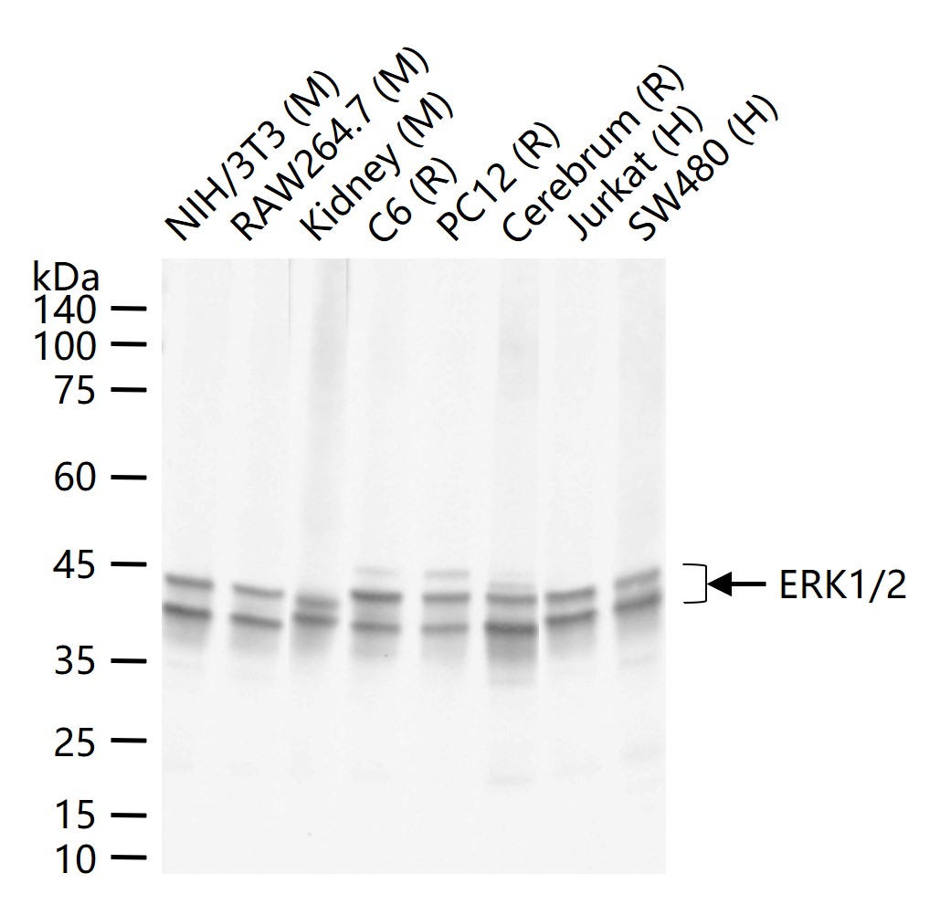
25 ug total protein per lane of various lysates (see on figure) probed with ERK1/2 monoclonal antibody, unconjugated (bsm-52259R) at 1:1000 dilution and 4°C overnight incubation. Followed by conjugated secondary antibody incubation at r.t. for 60 min.
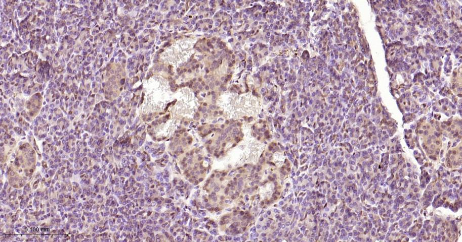
Paraformaldehyde-fixed, paraffin embedded Human Pancreas; Antigen retrieval by boiling in sodium citrate buffer (pH6.0) for 15 min; Antibody incubation with ERK1/2 Monoclonal Antibody, Unconjugated(bsm-52259R) at 1:200 overnight at 4°C, followed by conjugation to the bs-0295G-HRP and DAB (C-0010) staining.
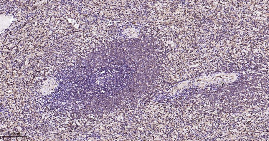
Paraformaldehyde-fixed, paraffin embedded Human Spleen; Antigen retrieval by boiling in sodium citrate buffer (pH6.0) for 15 min; Antibody incubation with ERK1/2 Monoclonal Antibody, Unconjugated(bsm-52259R) at 1:200 overnight at 4°C, followed by conjugation to the bs-0295G-HRP and DAB (C-0010) staining.
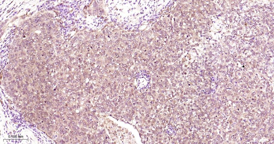
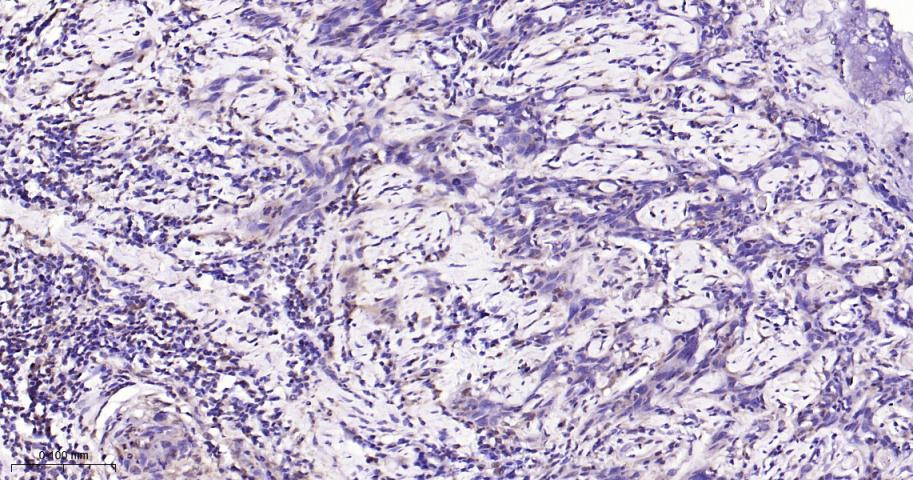
Paraformaldehyde-fixed, paraffin embedded Human Glioma; Antigen retrieval by boiling in sodium citrate buffer (pH6.0) for 15 min; Antibody incubation with ERK1/2 Monoclonal Antibody, Unconjugated(bsm-52259R) at 1:200 overnight at 4°C, followed by conjugation to the bs-0295G-HRP and DAB (C-0010) staining.
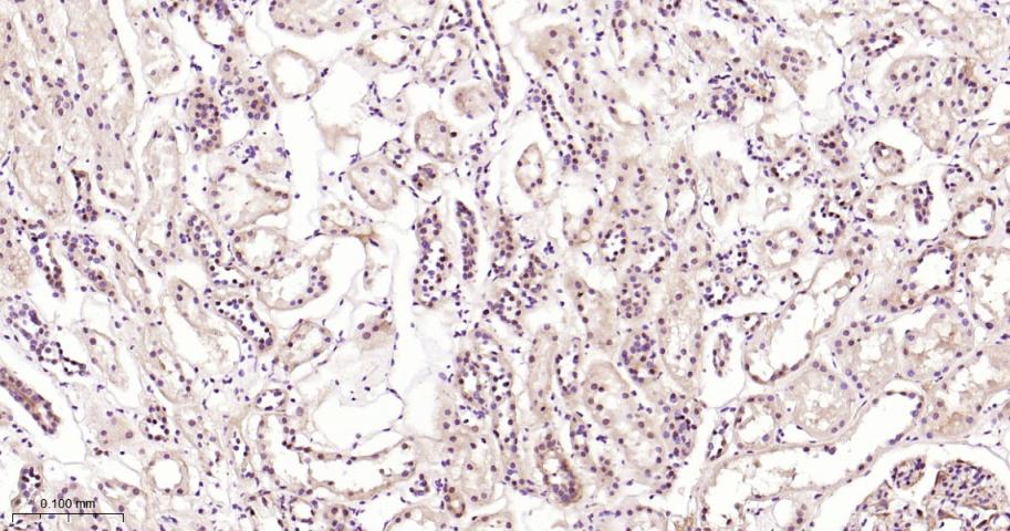
Paraformaldehyde-fixed, paraffin embedded Human Kidney; Antigen retrieval by boiling in sodium citrate buffer (pH6.0) for 15 min; Antibody incubation with ERK1/2 Monoclonal Antibody, Unconjugated(bsm-52259R) at 1:200 overnight at 4°C, followed by conjugation to the bs-0295G-HRP and DAB (C-0010) staining.
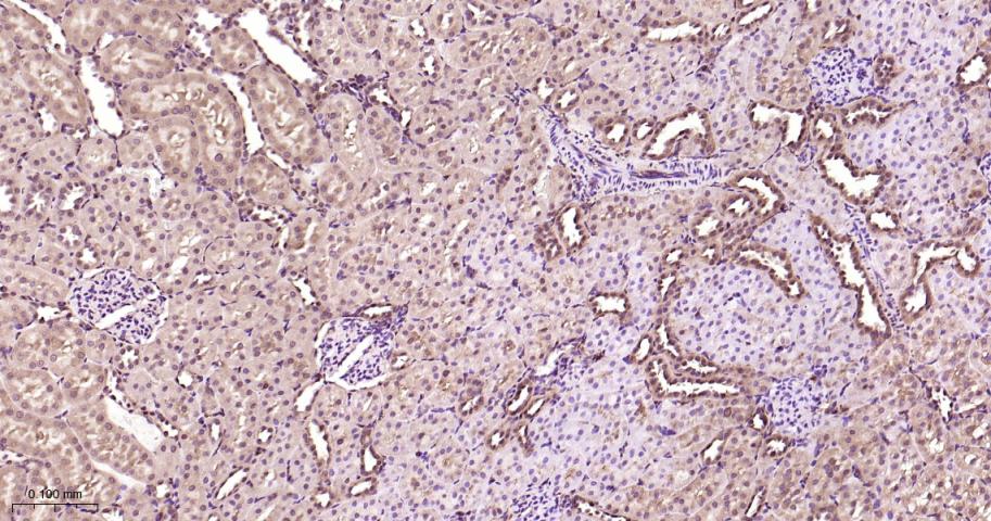
Paraformaldehyde-fixed, paraffin embedded Rat Kidney; Antigen retrieval by boiling in sodium citrate buffer (pH6.0) for 15 min; Antibody incubation with ERK1/2 Monoclonal Antibody, Unconjugated(bsm-52259R) at 1:200 overnight at 4°C, followed by conjugation to the bs-0295G-HRP and DAB (C-0010) staining.
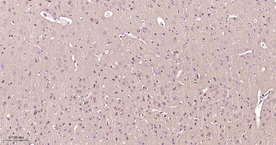
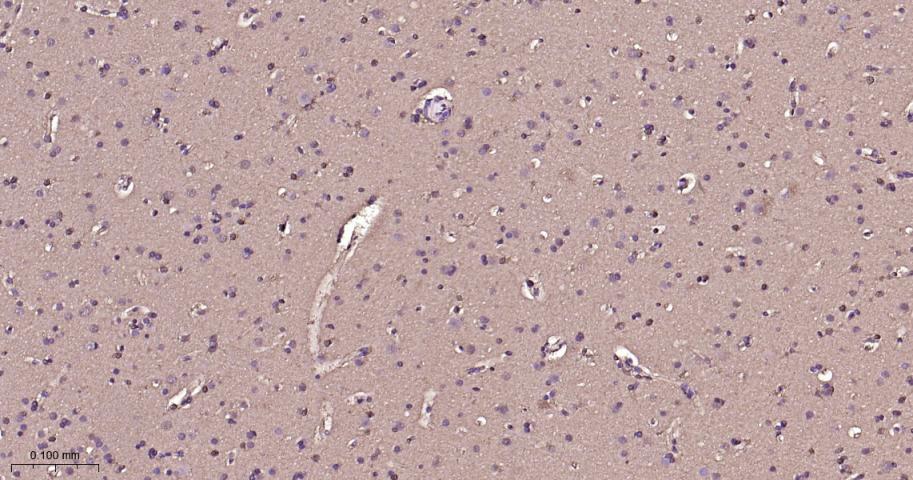
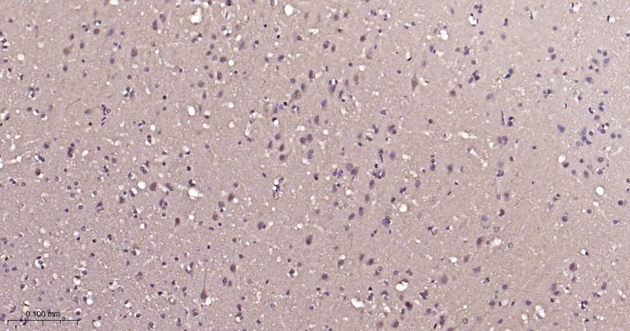
Paraformaldehyde-fixed, paraffin embedded Rat Cerebrum; Antigen retrieval by boiling in sodium citrate buffer (pH6.0) for 15 min; Antibody incubation with ERK1/2 Monoclonal Antibody, Unconjugated(bsm-52259R) at 1:200 overnight at 4°C, followed by conjugation to the bs-0295G-HRP and DAB (C-0010) staining.
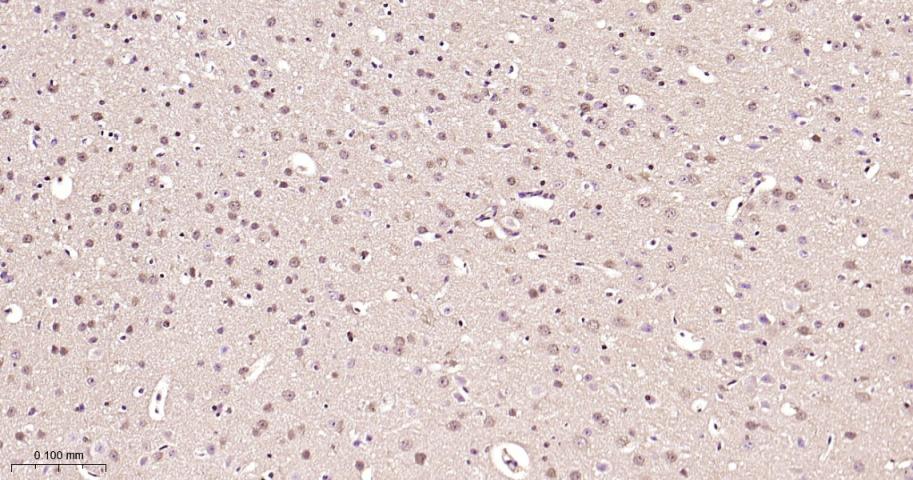
Paraformaldehyde-fixed, paraffin embedded Mouse Cerebrum; Antigen retrieval by boiling in sodium citrate buffer (pH6.0) for 15 min; Antibody incubation with ERK1/2 Monoclonal Antibody, Unconjugated(bsm-52259R) at 1:200 overnight at 4°C, followed by conjugation to the bs-0295G-HRP and DAB (C-0010) staining.
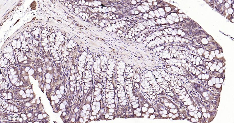
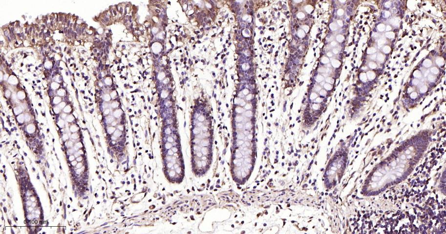
Paraformaldehyde-fixed, paraffin embedded Rat Colon; Antigen retrieval by boiling in sodium citrate buffer (pH6.0) for 15 min; Antibody incubation with ERK1/2 Monoclonal Antibody, Unconjugated(bsm-52259R) at 1:200 overnight at 4°C, followed by conjugation to the bs-0295G-HRP and DAB (C-0010) staining.
Paraformaldehyde-fixed, paraffin embedded Human Breast Cancer; Antigen retrieval by boiling in sodium citrate buffer (pH6.0) for 15 min; Antibody incubation with ERK1/2 Monoclonal Antibody, Unconjugated(bsm-52259R) at 1:200 overnight at 4°C, followed by conjugation to the SP Kit (Rabbit, SP-0023) and DAB (C-0010) staining.
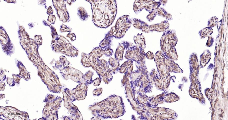
Paraformaldehyde-fixed, paraffin embedded Human Placenta; Antigen retrieval by boiling in sodium citrate buffer (pH6.0) for 15 min; Antibody incubation with ERK1/2 Monoclonal Antibody, Unconjugated(bsm-52259R) at 1:200 overnight at 4°C, followed by conjugation to the SP Kit (Rabbit, SP-0023) and DAB (C-0010) staining.
Paraformaldehyde-fixed, paraffin embedded Human Breast; Antigen retrieval by boiling in sodium citrate buffer (pH6.0) for 15 min; Antibody incubation with ERK1/2 Monoclonal Antibody, Unconjugated(bsm-52259R) at 1:200 overnight at 4°C, followed by conjugation to the SP Kit (Rabbit, SP-0023) and DAB (C-0010) staining.
Paraformaldehyde-fixed, paraffin embedded Human Cerebrum; Antigen retrieval by boiling in sodium citrate buffer (pH6.0) for 15 min; Antibody incubation with ERK1/2 Monoclonal Antibody, Unconjugated(bsm-52259R) at 1:200 overnight at 4°C, followed by conjugation to the SP Kit (Rabbit, SP-0023) and DAB (C-0010) staining.
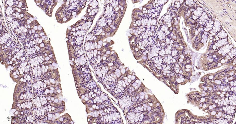
Paraformaldehyde-fixed, paraffin embedded Human Colon; Antigen retrieval by boiling in sodium citrate buffer (pH6.0) for 15 min; Antibody incubation with ERK1/2 Monoclonal Antibody, Unconjugated(bsm-52259R) at 1:200 overnight at 4°C, followed by conjugation to the SP Kit (Rabbit, SP-0023) and DAB (C-0010) staining.
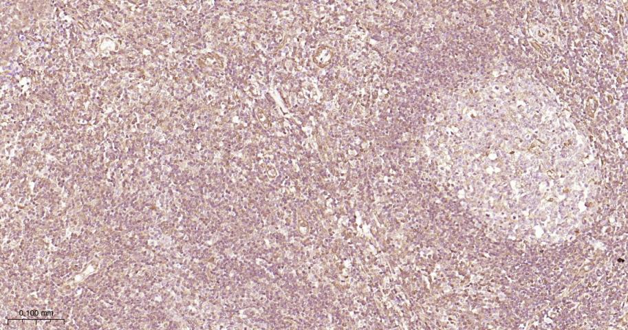
Paraformaldehyde-fixed, paraffin embedded Human Tonsil; Antigen retrieval by boiling in sodium citrate buffer (pH6.0) for 15 min; Antibody incubation with ERK1/2 Monoclonal Antibody, Unconjugated(bsm-52259R) at 1:200 overnight at 4°C, followed by conjugation to the SP Kit (Rabbit, SP-0023) and DAB (C-0010) staining.
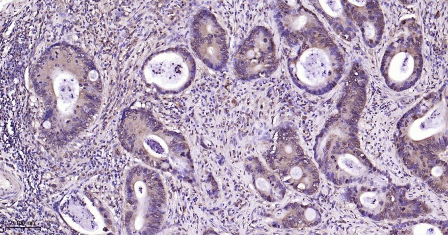
Paraformaldehyde-fixed, paraffin embedded Human Colon Cancer; Antigen retrieval by boiling in sodium citrate buffer (pH6.0) for 15 min; Antibody incubation with ERK1/2 Monoclonal Antibody, Unconjugated(bsm-52259R) at 1:200 overnight at 4°C, followed by conjugation to the SP Kit (Rabbit, SP-0023) and DAB (C-0010) staining.
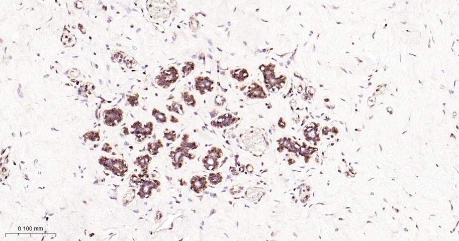
Paraformaldehyde-fixed, paraffin embedded Human Cervical Cancer; Antigen retrieval by boiling in sodium citrate buffer (pH6.0) for 15 min; Antibody incubation with ERK1/2 Monoclonal Antibody, Unconjugated(bsm-52259R) at 1:200 overnight at 4°C, followed by conjugation to the SP Kit (Rabbit, SP-0023) and DAB (C-0010) staining.
Paraformaldehyde-fixed, paraffin embedded Mouse Colon; Antigen retrieval by boiling in sodium citrate buffer (pH6.0) for 15 min; Antibody incubation with ERK1/2 Monoclonal Antibody, Unconjugated(bsm-52259R) at 1:200 overnight at 4°C, followed by conjugation to the SP Kit (Rabbit, SP-0023) and DAB (C-0010) staining.
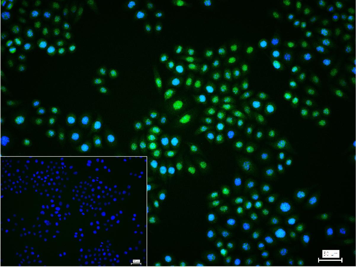
Tissue/cell:A549 cell;4% Paraformaldehyde-fixed;Triton X-100 at room temperature for 20 min; Blocking buffer (normal goat serum,C-0005) at 37°C for 20 min; Antibody incubation with (ERK1/2) monoclonal Antibody, Unconjugated (bsm-52259R) 1:100, 90 minutes at 37°C; followed by a FITC conjugated Goat Anti-Rabbit IgG antibody at 37°C for 90 minutes, DAPI (blue, C02-04002) was used to stain the cell nuclei.
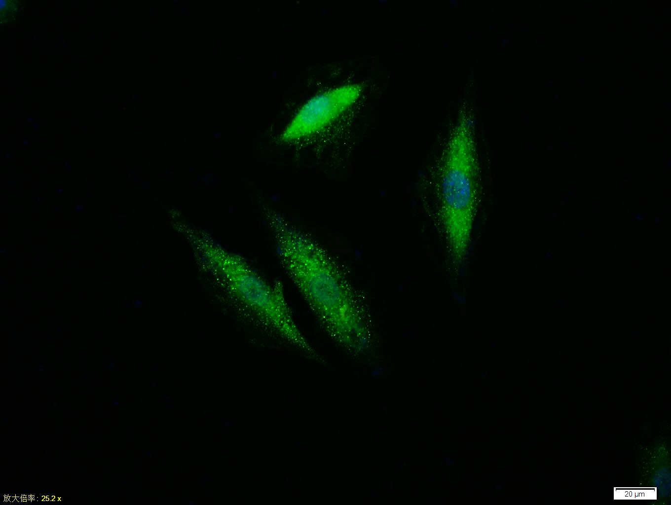
4% Paraformaldehyde-fixed Hela (H) cell; Triton X-100 at r.t. for 20 min; Antibody incubation with (ERK1/2) monoclonal Antibody, unconjugated (bsm-52259R) 1:100, 90 min at 37°C; followed by conjugated Goat Anti-Rabbit IgG antibody (green, bs-60295G-BF488) at 37°C for 90 min, DAPI (blue, C02-04002) was used to stain the cell nuclei. PBS instead of the primary antibody was used as the blank control.
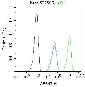
Blank control: Hela.
Primary Antibody (green line): Rabbit Anti-ERK1/2 antibody (bsm-52259R)
Dilution: 1μg /10^6 cells;
Isotype Control Antibody (orange line): Rabbit IgG .
Secondary Antibody : Goat anti-rabbit IgG-AF647
Dilution: 1μg /test.
Protocol
The cells were fixed with 4% PFA (10min at room temperature)and then permeabilized with 90% ice-cold methanol for 20 min at -20℃. The cells were then incubated in 5%BSA to block non-specific protein-protein interactions for 30 min at room temperature .Cells stained with Primary Antibody for 30 min at room temperature. The secondary antibody used for 40 min at room temperature. Acquisition of 20,000 events was performed.
|
| 1、抗体溶解方法 | |
| 2、抗体修复方式 | |
| 3、常用试剂的配制 | |
| 4、免疫组化操作步骤 | |
| 5、免疫组化问题解答 | |
| 6、Western Blotting 操作步骤 | |
| 7、Western Blotting 问题解答 | |
| 8、关于肽链的设计 | |
| 9、多肽的溶解与保存 | |
| 10、酶标抗体效价测定程序 | |


