

| 产品编号 | bs-41257R |
| 英文名称 | PGP9.5 Rabbit pAb |
| 中文名称 | 神经细胞胞浆蛋白9.5/蛋白基因产物9.5抗体 |
| 别 名 | HEL-117; HEL-S-53; NDGOA; PARK5; PGP 9.5; PGP9.5; PGP95; SPG79; SPG79A; UCHL-1; Uch-L1; gad; UCHL1_HUMAN; UCHL1; Neuron cytoplasmic protein 9.5; PGP 9.5 (PGP9.5); Ubiquitin thioesterase L1; 3.4.19.12; UCHL1_MOUSE; UCHL1_PIG; UCHL1_RAT; |
| 研究领域 | 细胞生物 神经生物学 细胞类型标志物 泛素 |
| 抗体来源 | Rabbit |
| 克隆类型 | Polyclonal |
| 克 隆 号 | |
| 交叉反应 | Human,Mouse,Rat |
| 产品应用 | WB=1:500-2000,ICC/IF=1:50-200
not yet tested in other applications. optimal dilutions/concentrations should be determined by the end user. |
| 理论分子量 | 25 kDa |
| 检测分子量 | 26 |
| 细胞定位 | 细胞浆 细胞膜 |
| 性 状 | Liquid |
| 浓 度 | 1mg/ml |
| 免 疫 原 | Recombinant human UCHL1 |
| 亚 型 | IgG |
| 纯化方法 | affinity purified by Protein A |
| 缓 冲 液 | 0.01M TBS (pH7.4) with 1% BSA, 0.02% Proclin300 and 50% Glycerol. |
| 保存条件 | Store at -20℃ for one year. Avoid repeated freeze/thaw cycles. The lyophilized antibody is stable at room temperature for at least one month and for greater than a year when kept at -20°C. When reconstituted in sterile pH 7.4 0.01M PBS or diluent of antib |
| 注意事项 | This product as supplied is intended for research use only, not for use in human, therapeutic or diagnostic applications. |
| PubMed | PubMed |
| 产品介绍 |
The protein encoded by this gene belongs to the peptidase C12 family. This enzyme is a thiol protease that hydrolyzes a peptide bond at the C-terminal glycine of ubiquitin. This gene is specifically expressed in the neurons and in cells of the diffuse neuroendocrine system. Mutations in this gene may be associated with Parkinson disease.[provided by RefSeq, Sep 2009] Function: Ubiquitin-protein hydrolase involved both in the processing of ubiquitin precursors and of ubiquitinated proteins. This enzyme is a thiol protease that recognizes and hydrolyzes a peptide bond at the C-terminal glycine of ubiquitin. Also binds to free monoubiquitin and may prevent its degradation in lysosomes. The homodimer may have ATP-independent ubiquitin ligase activity. Subcellular Location: Cytoplasm. Endoplasmic reticulum membrane. About 30% of total UCHL1 is associated with membranes in brain. Tissue Specificity: Found in neuronal cell bodies and processes throughout the neocortex (at protein level). Expressed in neurons and cells of the diffuse neuroendocrine system and their tumors. Weakly expressed in ovary. Down-regulated in brains from Parkinson disease and Alzheimer disease patients. Post-translational modifications: O-glycosylated. DISEASE: Defects in UCHL1 are the cause of Parkinson disease type 5 (PARK5) [MIM:613643]; also known as Parkinson disease autosomal dominant 5. PARK5 is a complex neurodegenerative disorder with manifestations ranging from typical Parkinson disease to dementia with Lewy bodies. Clinical features include parkinsonian symptoms (resting tremor, rigidity, postural instability and bradykinesia), dementia, diffuse Lewy body pathology, autonomic dysfunction, hallucinations and paranoia. Similarity: Belongs to the peptidase C12 family. SWISS: P09936 Gene ID: 7345 Database links: Entrez Gene: 7345 Human Entrez Gene: 22223 Mouse Entrez Gene: 101117250 Sheep Entrez Gene: 325119 Zebrafish Omim: 191342 Human SwissProt: P09936 Human SwissProt: Q9R0P9 Mouse Unigene: 518731 Human Unigene: 29807 Mouse Unigene: 107213 Rat |
| 产品图片 |
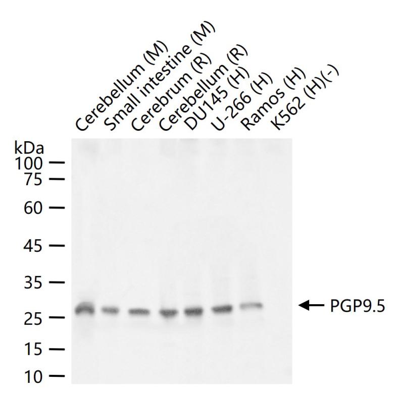
25 ug total protein per lane of various lysates (see on figure) probed with PGP9.5 polyclonal antibody, unconjugated (bs-41257R) at 1:1000 dilution and 4°C overnight incubation. Followed by conjugated secondary antibody incubation at r.t. for 60 min.
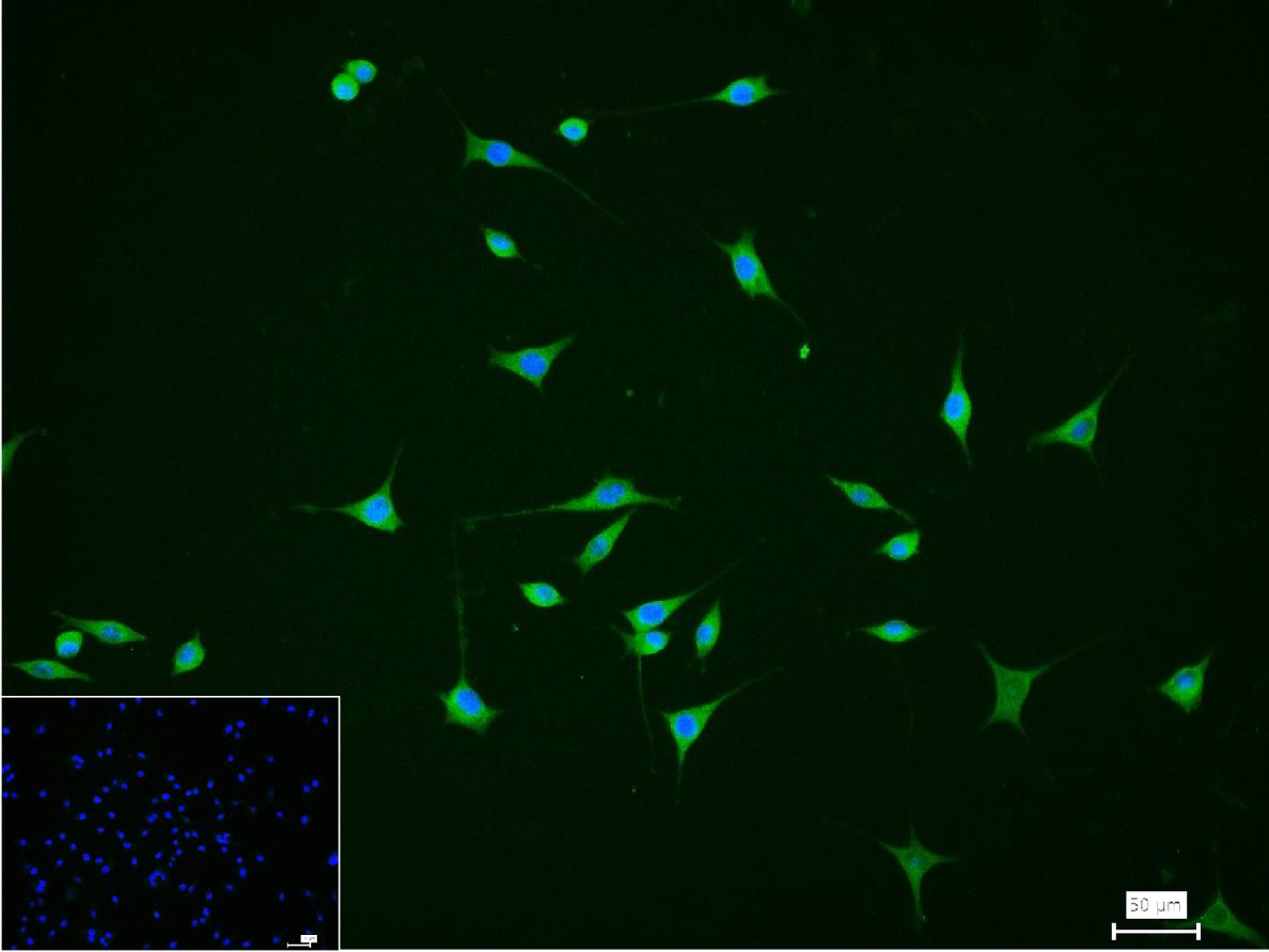
4% Paraformaldehyde-fixed SH-SY5Y (H) cell; Triton X-100 at r.t. for 20 min; Antibody incubation with (PGP9.5) polyclonal Antibody, unconjugated (bs-41257R) 1:100, 90 min at 37°C; followed by conjugated Goat Anti-Rabbit IgG antibody (green, bs-40295G-FITC) at 37°C for 90 min, DAPI (blue, C02-04002) was used to stain the cell nuclei. PBS instead of the primary antibody was used as the blank control.
|
| 1、抗体溶解方法 | |
| 2、抗体修复方式 | |
| 3、常用试剂的配制 | |
| 4、免疫组化操作步骤 | |
| 5、免疫组化问题解答 | |
| 6、Western Blotting 操作步骤 | |
| 7、Western Blotting 问题解答 | |
| 8、关于肽链的设计 | |
| 9、多肽的溶解与保存 | |
| 10、酶标抗体效价测定程序 | |


