
| 产品编号 | bsm-54037R |
| 英文名称 | PDPK1 Recombinant Rabbit mAb |
| 中文名称 | PDK1重组兔单抗 |
| 别 名 | PDK1; PDPK2; PDPK2P; PRO0461; PDPK1_HUMAN; PDPK1; hPDK1; 2.7.11.1; PDPK1_MOUSE; mPDK1; PDPK1_RAT; Protein kinase B kinase (PkB kinase); 3磷酸肌醇依赖性蛋白激酶1 |
| 研究领域 | 细胞生物 神经生物学 信号转导 细胞凋亡 激酶和磷酸酶 |
| 抗体来源 | Rabbit |
| 克隆类型 | Recombinant |
| 克 隆 号 | 2F9 |
| 交叉反应 | Human,Mouse,Rat |
| 产品应用 | WB=1:500-2000,IHC-P=1:500-2000,IHC-F=1:500-2000,IF=1:500-2000,Flow-Cyt=1:50-100
not yet tested in other applications. optimal dilutions/concentrations should be determined by the end user. |
| 理论分子量 | 61 kDa |
| 细胞定位 | 细胞浆 细胞膜 |
| 性 状 | Liquid |
| 浓 度 | 1mg/ml |
| 免 疫 原 | KLH conjugated synthetic peptide derived from human PDK1 |
| 亚 型 | IgG |
| 纯化方法 | affinity purified by Protein A |
| 缓 冲 液 | 0.01M TBS (pH7.4) with 1% BSA, 0.02% Proclin300 and 50% Glycerol. |
| 保存条件 | Shipped at 4℃. Store at -20℃ for one year. Avoid repeated freeze/thaw cycles. |
| 注意事项 | This product as supplied is intended for research use only, not for use in human, therapeutic or diagnostic applications. |
| PubMed | PubMed |
| 产品介绍 |
PDK1 (3 Phosphoinositide Dependent Protein Kinase 1) phosphorylates AGC kinases. PDK1 activates conventional PKC and PKC zeta through phosphorylation of critical threonine residues in the activation loop. PDK1 also phosphorylates Protein Kinase B (PKB) at threonine 308 in the presence of phosphatidylinositol-3,4,5-trisphosphate. Active Akt inactivates Glycogen Synthase Kinase 3 (GSK3), eventually leading to the dephosphorylation and activation of glycogen synthase and the stimulation of glycogen synthesis. Because of the role that PDK plays in insulin-induced glycogen synthesis and PKC activation it is a potentially important target for metabolic drug research. There are three named isoforms. Function: Phosphorylates and activates not only PKB/AKT, but also PKA, PKC-zeta, RPS6KA1 and RPS6KB1. May play a general role in signaling processes and in development (By similarity). Isoform 3 is catalytically inactive. Subunit: Interacts with NPRL2. Subcellular Location: Cytoplasm. Membrane; Peripheral membrane protein. Note=Membrane-associated after cell stimulation leading to its translocation. Tyrosine phosphorylation seems to occur only at the plasma membrane. Tissue Specificity: Appears to be expressed ubiquitously. Post-translational modifications: Phosphorylated on tyrosine and serine/threonine. Phosphorylation on Ser-241 in the activation loop is required for full activity. PDK1 itself can autophosphorylate Ser-241, leading to its own activation. Similarity: Belongs to the protein kinase superfamily. AGC Ser/Thr protein kinase family. PDK1 subfamily. Contains 1 PH domain. Contains 1 protein kinase domain. SWISS: O15530 Gene ID: 5170 Database links: Entrez Gene: 5170 Human Entrez Gene: 18607 Mouse Omim: 605213 Human SwissProt: O15530 Human SwissProt: Q9Z2A0 Mouse Unigene: 459691 Human Unigene: 10504 Mouse Unigene: 10905 Rat |
| 产品图片 |
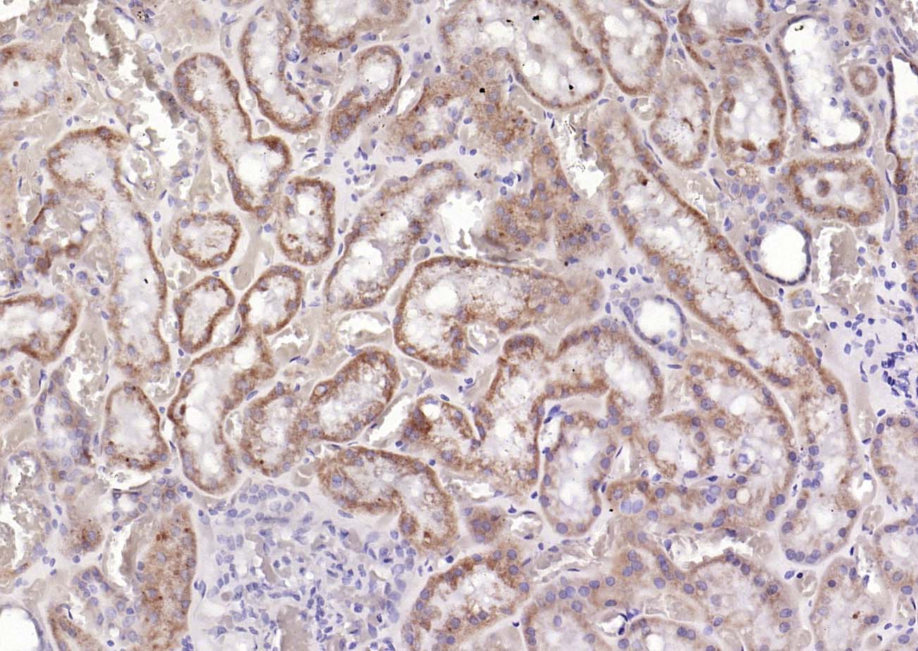
Paraformaldehyde-fixed, paraffin embedded (Human kidney); Antigen retrieval by boiling in sodium citrate buffer (pH6.0) for 15min; Block endogenous peroxidase by 3% hydrogen peroxide for 20 minutes; Blocking buffer (normal goat serum) at 37°C for 30min; Antibody incubation with (PDK1) Monoclonal Antibody, Unconjugated (bsm-54037R) at 1:200 overnight at 4°C, followed by operating according to SP Kit(Rabbit) (sp-0023) instructionsand DAB staining.
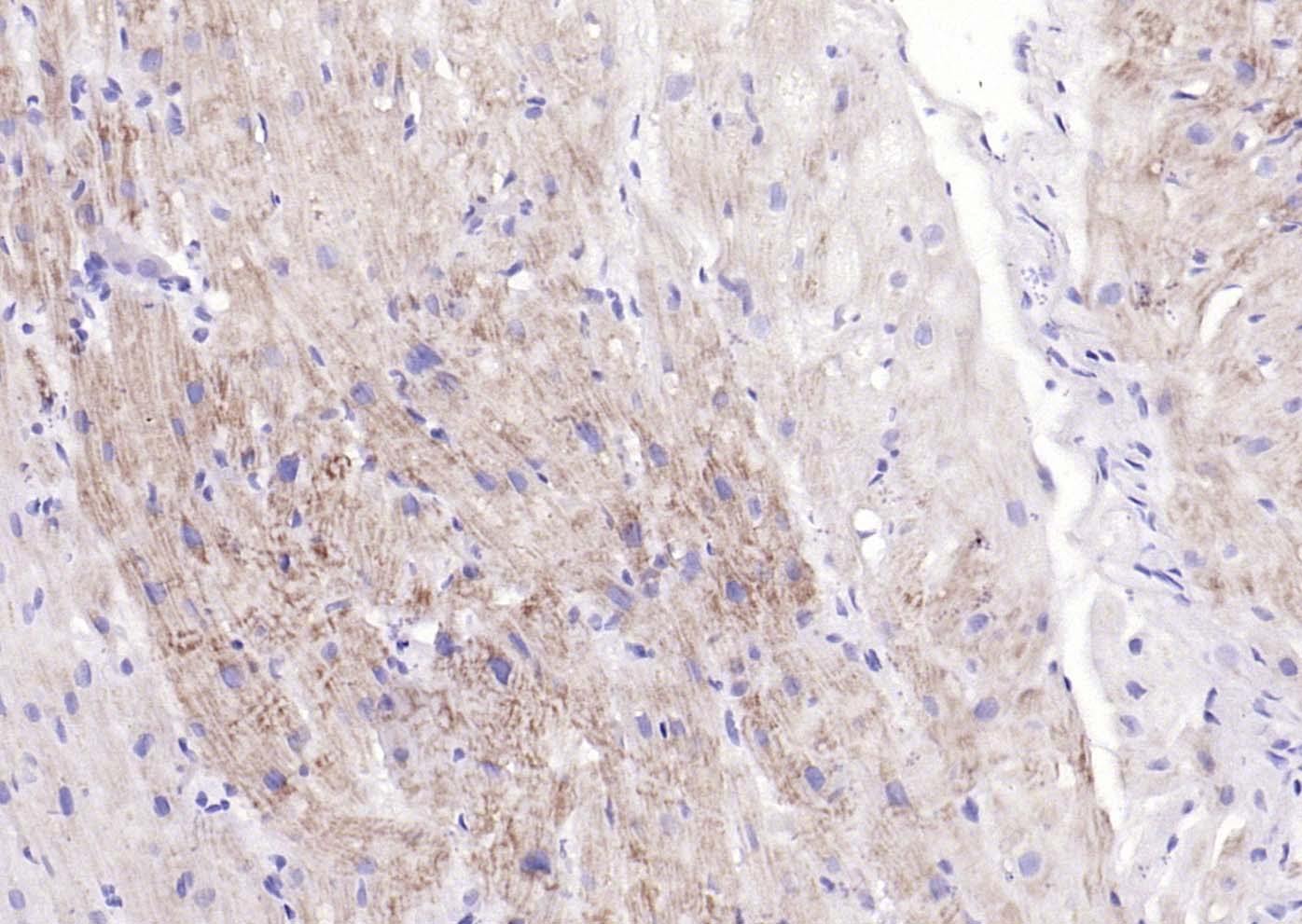
Paraformaldehyde-fixed, paraffin embedded (human myocardium); Antigen retrieval by boiling in sodium citrate buffer (pH6.0) for 15min; Block endogenous peroxidase by 3% hydrogen peroxide for 20 minutes; Blocking buffer (normal goat serum) at 37°C for 30min; Antibody incubation with (PDK1) Monoclonal Antibody, Unconjugated (bsm-54037R) at 1:200 overnight at 4°C, followed by operating according to SP Kit(Rabbit) (sp-0023) instructionsand DAB staining.
|
| 1、抗体溶解方法 | |
| 2、抗体修复方式 | |
| 3、常用试剂的配制 | |
| 4、免疫组化操作步骤 | |
| 5、免疫组化问题解答 | |
| 6、Western Blotting 操作步骤 | |
| 7、Western Blotting 问题解答 | |
| 8、关于肽链的设计 | |
| 9、多肽的溶解与保存 | |
| 10、酶标抗体效价测定程序 | |


