
| 产品编号 | bsm-52647R |
| 英文名称 | Tissue factor Recombinant Rabbit mAb |
| 中文名称 | 组织因子(CD142)兔单克隆抗体 |
| 别 名 | TF_HUMAN; F3; TF; Coagulation factor III; Thromboplastin; TF_MOUSE; Cf-3; Cf3; TF_RAT; |
 |
Specific References (1) | bsm-52647R has been referenced in 1 publications.
[IF=5.118] Masahiro Terasawa. et al. Anti-Inflammatory Activity of Orally Administered Monostroma nitidum Rhamnan Sulfate against Lipopolysaccharide-Induced Damage to Mouse Organs and Vascular Endothelium. Mar Drugs. 2022 Feb;20(2):121 WB ; Mouse.
|
| 研究领域 | 心血管 细胞生物 免疫学 生长因子和激素 细胞表面分子 |
| 抗体来源 | Rabbit |
| 克隆类型 | Recombinant |
| 克 隆 号 | 6A4 |
| 交叉反应 | Human,Mouse,Rat |
| 产品应用 | WB=1:500-2000,IHC-P=1:100-500,IHC-F=1:100-500,IF=1:100-500,ICC/IF=1:50-200
not yet tested in other applications. optimal dilutions/concentrations should be determined by the end user. |
| 理论分子量 | 35 kDa |
| 检测分子量 | 47 |
| 细胞定位 | 细胞膜 |
| 性 状 | Liquid |
| 浓 度 | 1mg/ml |
| 免 疫 原 | KLH conjugated synthetic peptide derived from human Tissue factor: 1-100/295 |
| 亚 型 | IgG |
| 纯化方法 | affinity purified by Protein A |
| 缓 冲 液 | 0.01M TBS (pH7.4) with 1% BSA, 0.02% Proclin300 and 50% Glycerol. |
| 保存条件 | Shipped at 4℃. Store at -20℃ for one year. Avoid repeated freeze/thaw cycles. |
| 注意事项 | This product as supplied is intended for research use only, not for use in human, therapeutic or diagnostic applications. |
| PubMed | PubMed |
| 产品介绍 |
This gene encodes coagulation factor III which is a cell surface glycoprotein. This factor enables cells to initiate the blood coagulation cascades, and it functions as the high-affinity receptor for the coagulation factor VII. The resulting complex provides a catalytic event that is responsible for initiation of the coagulation protease cascades by specific limited proteolysis. Unlike the other cofactors of these protease cascades, which circulate as nonfunctional precursors, this factor is a potent initiator that is fully functional when expressed on cell surfaces. There are 3 distinct domains of this factor: extracellular, transmembrane, and cytoplasmic. This protein is the only one in the coagulation pathway for which a congenital deficiency has not been described. Alternate splicing results in multiple transcript variants.[provided by RefSeq, May 2010] Function: Initiates blood coagulation by forming a complex with circulating factor VII or VIIa. The [TF:VIIa] complex activates factors IX or X by specific limited protolysis. TF plays a role in normal hemostasis by initiating the cell-surface assembly and propagation of the coagulation protease cascade. Subunit: Interacts with HSPE; the interaction, inhibited by heparin, promotes the generation of activated factor X and activates coagulation in the presence of activated factor VII. Subcellular Location: Isoform 1: Membrane; Single-pass type I membrane protein. Isoform 2: Secreted. Tissue Specificity: Lung, placenta and pancreas. Similarity: Belongs to the tissue factor family. SWISS: P13726 Gene ID: 2152 Database links: Entrez Gene: 2152 Human Entrez Gene: 14066 Mouse Omim: 134390 Human SwissProt: P13726 Human SwissProt: P20352 Mouse Unigene: 62192 Human Unigene: 273188 Mouse Unigene: 9980 Rat |
| 产品图片 |
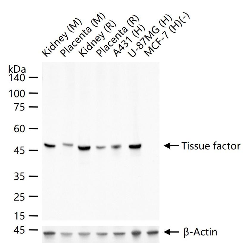
25 ug total protein per lane of various lysates (see on figure) probed with Tissue factor monoclonal antibody, unconjugated (bsm-52647R) at 1:1000 dilution and 4°C overnight incubation. Followed by conjugated secondary antibody incubation at r.t. for 60 min.
|
| 1、抗体溶解方法 | |
| 2、抗体修复方式 | |
| 3、常用试剂的配制 | |
| 4、免疫组化操作步骤 | |
| 5、免疫组化问题解答 | |
| 6、Western Blotting 操作步骤 | |
| 7、Western Blotting 问题解答 | |
| 8、关于肽链的设计 | |
| 9、多肽的溶解与保存 | |
| 10、酶标抗体效价测定程序 | |


